

Abstract
OBJECTIVE Hepatocellular carcinoma (HCC) is a hypervascular tumor for which angiogenesis plays an important role in its progression. The aim of this study was to investigate the expression of TWIST and VEGF and determine their roles in angiogenesis of HCC.
METHODS Expression Twist and VEGF mRNA was determined by realtime RT-PCR in 30 pairs of hepatocellular carcinoma and matched noncancerous tissues. Immunohistochemistry was carried out to analyze the protein expression of Twist and VEGF in 40 hepatocellular carcinoma cases. Staining of endothelial cells for CD34 was used to evaluate the microvessel density (MVD).
RESULTS We found that the HCC specimens showing positive Twist expression in tumor cells had a higher microvessel density than those without Twist expression. Furthermore, we found that overexpression of the Twist protein positively correlated with up-regulation of VEGF in the HCC tissues (r=0.479, P=0,002).
CONCLUSION Our results demonstrate that Twist may play an important role in the angiogenesis of HCC and a high-level of Twist expression may be related to the malignant potential of tumor cells.
keywords
Hepatocellular carcinoma (HCC) has become a worldwide common malignant tumor which shows relatively poor prognosis and rapid progression. Identification ofthe key molecular targets related to hepatocarcinogenesis has significant therapeutic implications. However, the genetic and epigenetic events of hepatocarcinogenesis are relatively understood.[1] TWIST, a basic helix-loop-helix (bHLH) transcription factor, can regulate mesodermal development and promote tumor cell metastasis. Recent studies have shown that Twist overexpression is able to induce angiogenesis, inhibit apoptosis, and trigger epithelial-mesenchymal transition that is pivotal for the transformation into an aggressive cancer phenotype.[2]
Angiogenesis is crucial for tumor growth and metastasis,[3] and there is evidence that solid tumors do not grow beyond 2~3 mm3 in volume without a new blood supply)[4] HCC is a typical hypervascular tumor. Angiogenesis, which is regulated by a balance between pro and antiangiogenic molecules is believed to contribute to malignant biological characteristics such as invasion and high rates of recurrence and metastasis.[5, 6] VEGF is the most angiogenic factor that promotes growth, proliferation and migration of endothelial cells[7] and in MCF-7 cells overexpressing Twist, an important regulator of tumor metastasis, VEGF synthesis is up-regulated. Furthermore, tumor xenografts of Twist overexpressed MCF-7 cells have a significantly elevated vascular volume and vascular permeability compared with MCF-7/vector control tumors,[2] indicating that Twist plays an important role in tumor angiogenesis. However, it is unclear whether Twist expression contributes to the neovascularization of human HCC. So our evaluation of the role of Twist in angiogenesis of human HCC is the first report to address this problem.
In this study, we examined the expression level of Twist mRNA and protein, and analyzed the relationship between Twist expression and angiogenesis in HCC tissues. Furthermore we evaluated the clinicopathological significance of Twist, VEGF and microvessel density (MVD) in patients with HCC.
Materials and Methods
Tissue samples
Samples of hepatocellular carcinoma tissues from 40 patients who had undergone surgery for HCC were obtained from the surgical department of the Tianjin Cancer Hospital. Written informed consent was obtained from each patient before tissue acquisition. HCC specimens were fixed in 10% buffered formalin and embedded in paraffin for immunohistochemistry. Among them, 30 pairs of hepatocellular carcinoma and matched non-cancerous tissues were collected and immediately frozen in liquid nitrogen and then stored at -80 °C before RNA extraction. The patients had a median age of 53 years (range 35~80 years).
RNA extraction and reverse transcripion
Total RNA was extracted and purified from 30 paired samples of fresh frozen cancerous tissues and non-cancerous liver tissues using the Trizol Reagent (Invitrogen, USA) following the manufacturer’s instructions. RNA from each sample was added to 20 ¼l of reaction mixture, and cDNA was synthesized with MML-V reverse transcriptase (TaKaRa, Dalian, China) according to the manufacturer’s instructions.
Real-time PCR
Real-time PCR analyses for Twist, VEGF and GAPDH mRNAs were performed using the ABI PRISM 5700 Sequence Detection System instrument and software. Primers of each gene were as follows: GAPDH: 5’-GAA GGT GAA GGT CGG AGT C-3’, 5’-GAA GAT GGT GAT GGG ATT TC-3’; Twist: 5’-GGG AGT CCG CAG TCT TAC GA-3’, 5’-AGA CCG A-GAAGG CGT AGC TG-3’; VEGF: 5’-GAA GGA GGA GGG CAG AAT CAT CAC-3’, 5’-CAC AGG ATG GCT TGA AGA TGT ACT C-3’.
An amplified GAPDH fragment was used as an internal quantitative control. Real-time PCR was performed with the SYBR Premix Ex Taqtm (TaKaRa, Dalian, China). Negative controls (cDNA-free solutions) were included in each reaction. The conditions were as follows: enzyme activation at 95 °C for 10 s for 1 cycle, followed by 40 cycles of denaturing at 95 °C for 5 s, annealing at 59 °C for 15s, and extension at 72 °C for 15 s. Data analysis using the 2-△△Ct method as follows: △△Ct= (Cttarget —Ctactin) tumour — (Cttarget — Ctactin)non — tumorous tissue.[8] Changes in expression levels of >2 were defined as up-or down-regulation.
Immunohistochemical analysis
Immunohistochemistry was performed to study altered protein expression in 40 human hepatocellular carcinoma tissues. Briefly, 4-¼m sections of routine formalin-fixed and paraffin-embedded material were de-paraffinized with xylene and rehydrated in graded alcohols and distilled water. Antigen retrieval was carried out by microwave treatment of the slides in sodium citrate buffer (pH 6.0) for 20 min. To quench the endo genous peroxidase activity, the sections were treated with 3% hydrogen peroxide in methanol for 10 min. After blocking the non-specific binding with 10% normal goat serum for 15 min, the sections were stained with rabbit monoclonal antibodies against Twist (1:50, Santa Cruz, USA), and mouse monoclonal antibodies for VEGF and CD34 (ZhongShan, China). The sections were incubated with atibodies over night at 4 °C and then washed with buffer. After washing, the tissue sections were then reacted with the biotinylated secondary antibody for 15 min and finally incubated with peroxidase-conjugated streptavidin at room temperature for 15 min. Color was developed with 3,3’-diaminobenzidine tetrachloride, and all slides were counterstained with hematoxylin. For negative controls, the primary antibodies were replaced with normal goat serum.
Evaluation of staining
For evaluation of Twist and VEGF expression, extent of staining was scored as 0 (0%), 1 (l%~25%), 2 (26%~50%), 3 (51%~75%), and 4 (76%~100%) according to the percentage of the positive staining areas in relation to the whole cancer areas. Staining intensity was scored as 0 (negative), 1 (weak), 2 (medium), and 3 (strong).The sum of the intensity and extent score was used as the final staining score (07) for Twist and VEGF. Tumors having a final staining score of >2 were considered to be positive.[9, 10]
Microvessel quantitation
Staining of endothelial cells for CD34 was used to e-valuate the MVD. Any CD34 positive endothelial cell clusters clearly separated from each other were considered as single countable microvessels. Larger vessels with muscular walls were excluded from counting. In each sample five areas of most prominent vascular density (hot spots) were identified at x40 power field and microvessels were counted under x200 magnification. The mean count was used for data analysis. The results were assessed by 2 observers who were unaware of any details regarding the patient background.
Statistical analysis
Independent-sample T test and one-way ANOVA was used to compare the mean MVD counts of groups of patients. The χ2 test was used to analyze the relationship between the protein expression and clinicopathologic characteristis. A bivariate Spearman test was chosen to assess the correlation between relative gene expression levels. Results were considered significant when P<0.05 was obtained. All the statistical analyses were performed using SPSS 11.5 for Windows.
Results
The expression pattern of Twist and VEGF in HCC
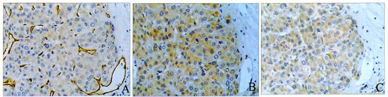
Real-time RT-PCR revealed that expression of Twist messenger RNA in the HCC tissues was increased in 73% (22/30) of cases compared to non-cancerous tissues. Up-regulated VEGF mRNA was detected in 67% (20/30) of the samples and 70% (14/20) of cases correlated with overexpression of Twist. Immumohistochemical analyses showed that Twist staining was in both the nuclei and cytoplasm, but cytoplasmic staining was predominant in most cases. The expression of Twist was positive in 55% (22/40) of the cases. Specific VEGF immunostaining, mostly in cytoplasm, was positively expressed in 62% (25/40) of the HCC patients (Fig.l).

Intensive staining of microvessels by anti-CD34 immunostaining (brownish staining) in a tumor section (A).Twist was highly expressed in the same patient(B).Staining of VEGF was also strong in the same patient(C). Photos were taken under 200⨯ magnifications.
Correlation among Twist, VEGF and MVD
We found that the mean MVD in cases of Twist-positive HCC specimens was higher than that in cases of Twist-negative HCC tissues although the difference failed to reach statistical significance (82.85±31.02 vs. 67.20±24.45; P=0.090). The mean MVD in the VEGF-positive group was significantly higher than the VEGF-negative group. (83.18+28.46 vs. 63.51 +26.36; P=0.036). Spearman correlation analysis showed there was a significant positive correlation between Twist protein expression and VEGF (r=0.479, P=0.002, Fig. 2).

Positive correlation between Twist and VEGF expression scorings in HCC tissue samples was observed by using the Spearman rank correlation coefficient.
The relationship between Twist, VEGF, MVD and dinicopathological features
The relationship between Twist, VEGF, MVD and dinicopathological features is summarized in Table 1.
The relationship between Twist,VEGF,MVD and clinicopathological features
There was no significant correlation between the expression of Twist protein and gender, tumor size, level of AFP or differentiation grade of HCC. However we found that positive expression of Twist was significantly associated with intrahepatic metastasis. There was no significant correlation between VEGF expression and any clinicopathological features. We also found that MVD in the tumors i 5 cm was higher than that in those >5 cm (85.88±25.98 vs. 66.59±29.64; P= 0.037).
Discussion
HCC is a highly malignant tumor with a propensity for vascular invasion and metastasis. Hepatic resection is the treatment of choice for HCC, but the prognosis after resection remains unsatisfactory because of a high incidence of recurrence related to tumor metastasis.[11] Tumor angiogenesis is an important determinant of invasiveness and progression of HCC. So it’s worthwhile to explore the role of mediators of angiogenesis in HCC.
As a novel oncogene, Twist has been studied in several types of tumors, including gastric, endometrial, prostate cancers and nasopharyngeal carcinoma. Overexpression of the Twist protein has been shown to correlate with tumor metastasis and poor outcome.[10, 12-14]
In this study, we found Twist mRNA as well as protein was up-regulated in HCC tissues in comparison with matched non-cancerous tissues. Further analysis suggested that overexpression of the Twist protein was positively correlated with intrahepatic metastasis. We also found an association between Twist expression and HCC angiogenesis, although this was not statistically significant. So Twist must play an important role in hepatic tumorigenesis and progression.
Growth, invasion and metastasis of malignant tumors depend on the process of angiogenesis. Twist has been recently recognized to be capable of inducing angiogenesis in vitro experiments and animal models. It has been observed that MCF-7 cells overexpressing Twist increased soluble VEGF expression by 10-fold when compared with empty vector control cells, and that MCF-7/Twist tumors exhibited higher vascular volume and vascular permeability than the MCF-7/vector control xenografts in a mouse model.[2] A recent study of human embryonic development also indicated that Twist was up-regulated during embryonic vascular formation. This indicates that Twist is not only a regulator of gastrulation, but also plays a part in angiogenesis during embryonic development.[15] However, the actual role of Twist in angiogenesis of HCC remains to be elucidated. In our present study, we found there was a trend towards a higher MVD in the tumors with positive Twist expression, although the difference was not statistically significant. Furthermore we observed a significant positive correlation between Twist protein expression and VEGF in HCC samples. These findings suggest that Twist promotes angiogenesis via up-regulation of VEGF synthesis in human FICC.
VEGF is the most angiogenic factor that can promote endothelial proliferation and increase vascular permeability by binding to its specific receptors in endothelial cells such as Fit-1, KDR/Flk-1 and Flt-4.[16] In our study, we found that VEGF mRNA was up-regulated in 67% (20/30) of the samples and VEGF protein was positively expressed in 62% of the HCC cases. With respect to the relationship between VEGF expression and clinicopathological factors, several previous studies have addressed this issue. Yamaguchi et al. [17] reported that VEGF was highly expressed in well-differentiated HCC and that the number of VEGF positive cases gradually decreased with tumor size. In another report, increased vascular endothelial growth factor expression was associated with tumor dedifferentiation.[18] In our present study, we failed to find any significant correlation between VEGF expression and the AEP level, tumor size, histological grade or metastasis. But we found that overexpression of VEGF significantly correlated with MVD. This indicated that VEGF may have important role in the angiogenesis of HCC. In addition, we found Twist and VEGF were both up-regulated in HCC samples. However, the exact molecular association remains unclear. Further study will be required to elucidate the relationship between the increases in Twist and VEGF expression in HCC.
The CD34 protein, an endothelial-specific marker, was immunohistochemically stained and MVD was determined in the HCC tissues in our study. We found that MVD in the Twist-positive tumors was higher than that in the Twist-negative tumors, although the difference was not statistically significant. We also found that MVD in the tumors less than 5 cm was significantly higher than the larger tumors. This result is in agreement with a previous study by Ng, et al.[19] In general, advanced HCCs are well supplied with arterial blood. In small HCCs, arteiy-like vessels are not well developed, capillarization of the blood spaces is present but in an incomplete form, and portal tracts are often present within cancerous nodules. These HCCs are thought to predominantly receive a portal blood supply. With the increase in tumor size, portal tracts decrease in number or disappear, and artery-like vessels gradually increase in number and size.[20] This may explain why the smaller tumors have more vasculature than those that are larger.
In conclusion, this study shows that the expression of Twist in HCC is related to tumor angiogenesis and invasiveness. In addition, Twist overexpression coincides with up-regulation of VEGF in HCC. These new findings may provide insight into a new therapeutic strategy for HCC by inhibiting Twist expression.
- Received October 18, 2006.
- Accepted November 17, 2006.
- Copyright © 2006 by Tianjin Medical University Cancer Institute & Hospital and Springer
References
In this issue


Jump to section
Related Articles
Cited By...
- No citing articles found.