

Abstract
Objective To explore the relationship of high S-phase kinase associated protein 2 (Skp2) expression with the characteristics of breast cancer.
Methods Using a immunohistochemical method, Skp2 expression was detected and evaluated in 30 normal tissues, 30 atypical ductal hyperplasias (ADH), 30 ductal carcinomas in situ (DCIS) and 56 Invasive carcinomas (including invasive specific carcinoma and invasive nonspecific carcinoma). The relationship between Skp2 expression and the characteristics of breast cancer was analyzed.
Results Skp2 expression varied among normal tissue, ADH, DCIS and invasive carcinomas (χ2 = 54.02, P<0.005). The positive level of Skp2 expression In DCIS was higher than that of normal tissue (χ2 = 21.82, P< 0.005) and there was a significant difference In Skp2 between DCIS and ADH (χ2=5.08, P<0.025) as well as between Invasive carcinomas and DCIS (χ2=6.52, P<0.01). The positive expression level of Skp2 in invasive carcinomas with positive lymph nodes was remarkably higher compared to those with negative lymph nodes. A significant difference in Skp2 expression was found between combined histological Grades I and II versus Grade III of invasive carcinomas (χ2=6.66, P<0.01).
Conclusion High Skp2 expression may play an important role in carcinogenesis and biological behavior of breast cancer.
keywords
In 1995 Skp2 was cloned by Zhang et al.[1] from human fibroblasts and it was shown that Skp2 interacts by combining with cyclin dependent kinase 2 (cyclin-CDK2). Skp2 is a member of the F-box family of substrate-recognition subunits of Skp2-Cullin-F-box ubiquitin-protein ligase complexes, and Skp2 has been implicated in the ubiquitin-mediated degradation of several key regulators of mammalian Gi progression, including the cyclin-ependent kinase inhibitor p27, a dosage-dependent tumor suppressor protein.
Since 1995, researchers have focused on Skp2 expression using immunohistochemical methods, gene knock-out animals and RT-PCR. Skp2 has been reported to be overexpressed in a variety of cancer types and to correlate with poor prognosis. It was found in a transgenic mouse line with high expression of Skp2 that these animals showed marked prostatic hyperplasia, dysplasia, and low-grade prostatic carcinoma.[2] In addition, amplification and high expression of Skp2 are associated with metastatic lymph nodes from non-small cell lung cancer.[3]
These studies indicated that Skp2 was related to the biological behavior of a variety of cancers, so we determined if there was a close relationship between Skp2 and breast cancer. The aim of our study was to explore the level of Skp2 expression using immunohistochemical methods in normal tissue, atypical ductal hyperplasia, ductal carcinoma in situ and invasive carcinoma (including invasive specific and nonspecific carcinoma). Our hope was that this research might offer a theoretical basis for diagnosis and therapy of breast cancer.
Materials And Methods
Materials
In this study, 146 tissues were examined, including 30 cases with normal tissues adjacent to an adenofibroma, 30 cases with ADH and 30 cases with DCIS. Invasive carcinomas included invasive specific carcinomas and invasive nonspecific carcinomas. In order to explore the relationship of axillary lymph nodes in breast cancer patients with Skp2, we selected 28 cases of invasive carcinoma with, 28 cases without métastasés of axillary lymph nodes. According to the degree of cell differentiation, the invasive carcinoma cases were separated into 3 grades including 2 cases of the Grade I (better differentiation), 32 cases of the Grade II (moderate differentiation), 22 cases of the Grade III (worse differentiation). The cases were diagnosed and classified according to "The WHO classification of tumors of the breast cancer and female genital organs".[4] All cases were female of ages 22 to 76 years with a median age of 49.
Methods
The 146 tissue samples were dehydrated, embedded and sectioned. All reagents, including mouse monoclonal antibody anti-Skp2, a PV-9000 kit and a DAB kit, were purchased from the ZhongShan Biotechnology Co. Ltd. A section of breast carcinoma with positive Skp2 expression was used as a positive control. Primary antibody was replaced by 1% bovine serum albumin as a negative control. The primary antibody was diluted to 1:100. All methods were performed strictly according to standard immunohistochemical procedures.
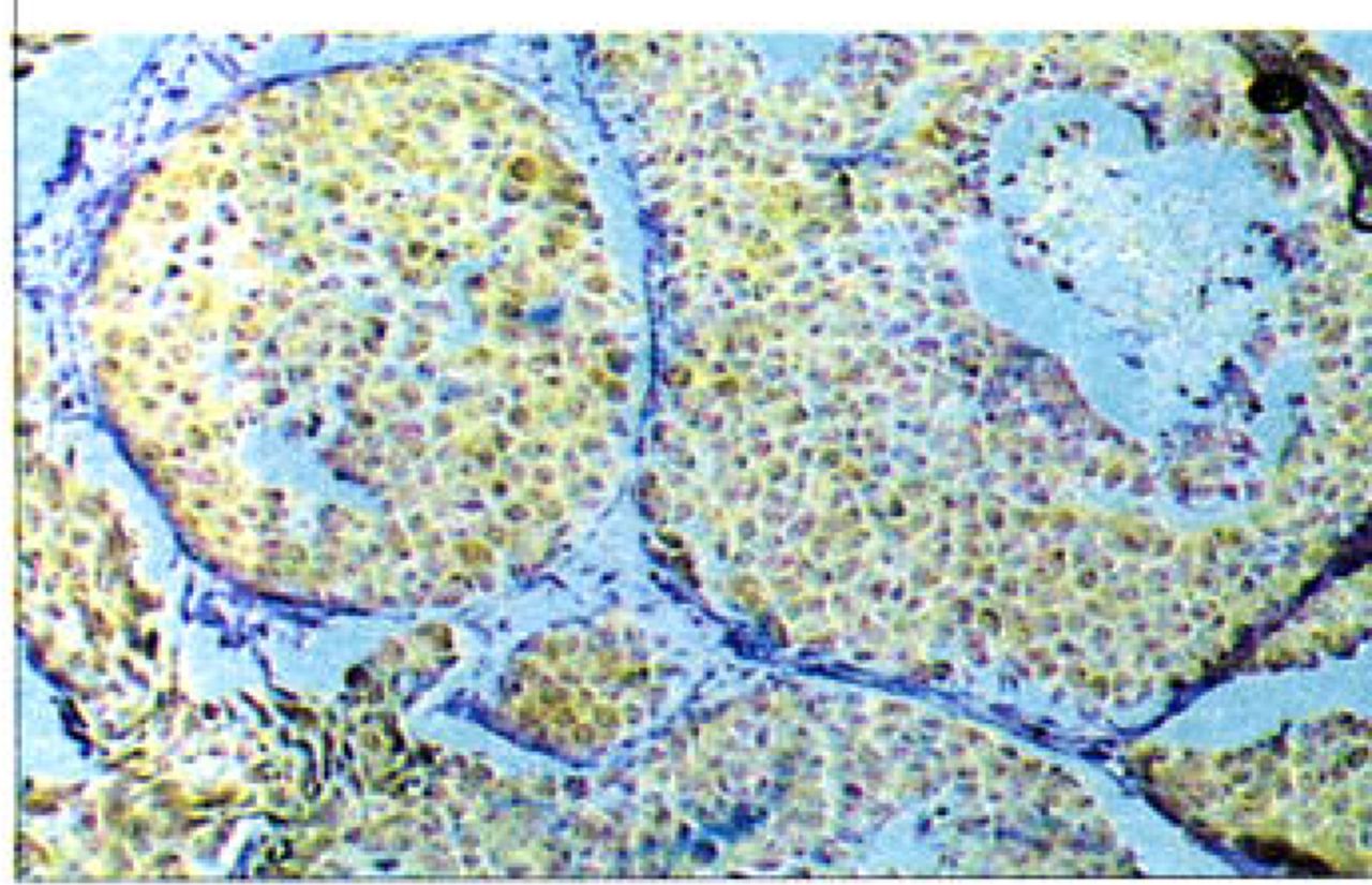
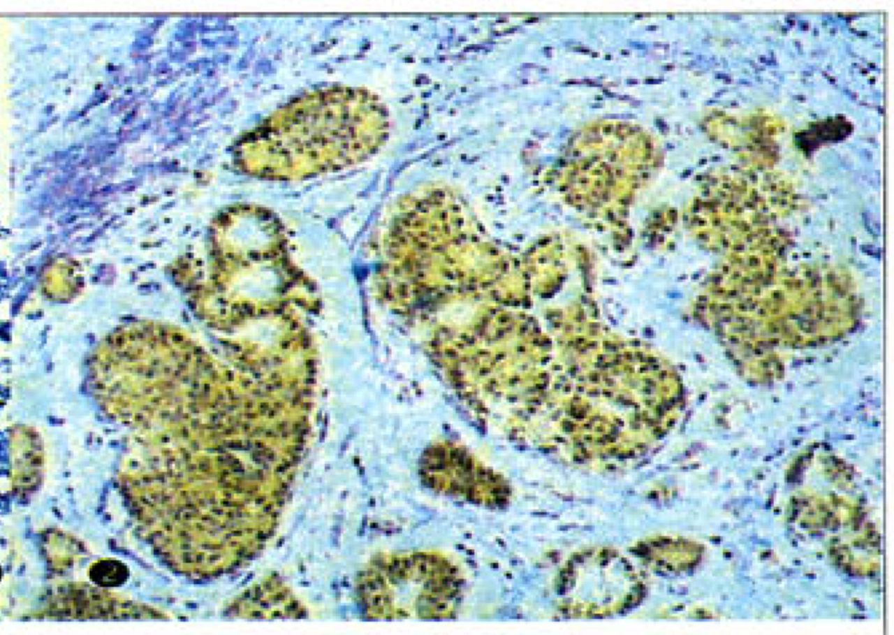
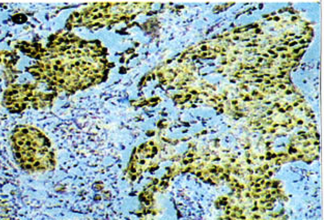
Amended Zheng et al.[5] immunohistochemical criteria of defining positive Skp2 cells was used to evaluate the results. The positive brown-yellow were particles located in the cytoplasm and nucleus. Cases defined as positive cases with high Skp2 expression had an average of >20% positive cells/10HP fields. Negative cases had lower Skp2 expression with <20%/10HP fields. The Skp2 positive ratio of each group was the number of Skp2 positive cases per total cases.
Statistical analysis
Statistical evaluation was performed with the y2 test. The Grade I and Grade II invasive carcinoma groups were combined in order to amplify sample size of these groups.
Results
Comparation of the rate of Skp2 positive expression among the four groups
Skp2 expression was negative in normal tissues Fig. 1 The percentage of positive cells was 65% in ADH Fig.2. Seventy five percent positive cells and 80% positive cells were seen respectively in DCIS and invasive carcinomas Fig.34 The positive ratio of Skp2 expression in the 4 groups is shown in Table 1. On the whole, there were differences in the 4 groups (χ2= 54.02, P<0.005). Skp2 expression in DCIS was significantly higher than that in normal tissues (χ2=21.82, P<0.005). A large difference was found between ADH and DCIS (χ2=5.08, P<0.025). Skp2 expression in DCIS was lower than that in invasive carcinomas (χ2=6.52,P<0.01, Table 1).
The positive rate of Skp2 expression in normal tissues, ADH, DCIS and invasive carcinoma (cases and percent)

Skp2 expression in nuclei and cytoplasm of normal tissues (Envision * 100).

Skp2 expression in nuclei and cytoplasm of ADH (Envision * 100).
Skp2 expression in nuclei and cytoplasm of DCIS (Envisionx 100).

Skp2 expression in nuclei and cytoplasm of invasive carcinoma ( Envisionx 100).
Correlation of Skp2 overexpression with histopafhological grade and with invasion of breast carcinoma
A significant difference was found in invasive carcinoma accompanied by positive axillary lymph nodes (ALN) compared to negative (χ2 = 7.38, P<0.025). Skp2 expression in the Grade I plus II of invasive carcinoma was lower than that in the Grade III of invasive carcinoma (χ26.66, P<0.01, Table 2).
The relationship between Skp2 expression and biological behavior of breast cancer
Discussion
Correlation of Skp2 with cell cycle control
In the cell cycle, the G1 to S transition is the key point that controls cell division. At present, two kinds of cell cycle regulators have been found to participate at this key point: one is a promoter, such as cyclin D/CDK2, cyclin E/CDK2 or p21wafl. Another is an inhibitor, such as p27kipl. Normally these two kinds of regulators not only depend on each other and but also inhibit each other to regulate normal cell division.
Skp2 is a member of the F-box family of substrate-recognition subunits of Skpl-Cullin-F-box(SCF) ubiquitin-protein ligase complexes. It is located in the nucleus and functions in the ubiquitin-mediated degradation of p27kipl, p21wafl, E2F, cyclin Dl, cyclin E, cyclin A and cyclin B.[6]
A novel isoform of Skp2, we named Skp2B, differs from Skp2 only in the C-terminal domain and unlike Skp2, localizes to the cytoplasm.m Skp2B is overexpressed as frequently as Skp2, and to higher levels than Skp2 in breast cancer cell lines and primary cancers. These findings therefore suggest that high expression of Skp2B may also be oncogenetic.
It has been confirmed that ubiquitin-mediated degradation of p27kipl is necessary for normal cell division.181 However, abnormal Skp2 gene copy numbers result in Skp2 overexpression, cyclin dependent kinase inhibitor (CKI) and p27kipl overdegradation, unlimited cell division and finally carcinogenesis. Skp2 overexpression has close relationship with carcinogenesis and its high expression has been found in many cancers, such those of the breast, stomach and lung.[5,9,10]
The correlation of high Skp2 expression and biologtical behavior of cancer
In our study the Skp2 positive level of DCIS was significantly higher than that in ADH, indicating that high Skp2 expression has a relationship with the malignancy of breast ductal epithelial cells (P<0.025). The mechanism of cell malignancy could be based on the SCF complex locating in the centrosome formed at cell division. One part of the SCF-Skp2 complex regulates centrosome duplication and chromosal replication. High Skp2 expression might lead to unlimited centrosome duplication,[11] aberrant cell division, multiploid and carcinoma.
It was found that Skp2 positive ratio in invasive carcinoma was significantly higher than that in DCIS (P<0.01). Furthermore, Skp2 expression in invasive carcinomas with positive lymph nodes was higher compared to carcinomas with negative lymph nodes (P<0.025). The results imply that high Skp2 expression is correlated with invasion and metastasis of breast carcinoma. Recently Yokoi et al.[12] and Li et al. [13] found that high Skp2 expression was associated with the metastasis of small cell lung cancer (SCLC) and colon carcinoma. The mechanism of metastasis might be related to the cellular microstructureal centrosome. It has been demonstrated that centrosome correlates with formation of fila pseudopods.[14] When Skp2 locates in the centrosome,[15] we supposed that high Skp2 expression in tumor cells might provive an ability for cells to migrate and form métastasés.
The Skp2 positive level of DCIS in our studies was strikingly higher than that of normal tissues, a finding which is consistent with reports from those outside China.[16] Therefore, more attention should be paid on the clinical significance of high Skp2 expression in breast cancer.
Our results showed that the level of Skp2 expression was significantly different in comparing Grade I and II of invasive carcinoma with the Grade III of invasive carcinoma, suggesting that there was a relationship between high Skp2 expression and ductal epithelial differentiation. This finding is consistant with Dowen’s results in cervical cancer.[17]
In addition, there is close relationship between Skp2 and the estrogen and progestin receptors, Her-2 and p27kipl. In 2002, Signoretti1'61 analyzed the correlation of high expression of Skp2 with the estrogen and progestin receptors using an immunohistochemical method, cell culture and oligonucleotide microarrays. They found that the positive ratio of Skp2 in ER'/PR samples was significantly higher than that of ER7PR+ tissues. Patients with ER'/PR’ are resistant to hormone therapy, so it can be supposed by this result that high expression of Skp2 in breast ductal epithelium was associated with poor prognosis. Foster et al.[18] suggested that estrogen down regulated p27kipl in breast cancer cells through Skp2. It has been proposed that high expression of the Skp2 protein in oral epithelium may lead to accelerated p27kipl proteolysis and contribute to malignant progression from dysplasia to oral epithelial carcinoma.[19] It was reported that an alternative reading frame protein (ARF),[20] as a tumor suppressor protein encoded by a gene located in the Ink4a/ARF gene locus, led to decreased levels of Cull and Skp2 and increased p27 stability in HER2/neu-overexpressing cells. As a consequence, it caused HER2/neuoverexpressing cells apoptosis. These results suggested that decreased Skp2 via ARF would have positive effect on therapy of HER2/neu-overexpressing patients.
Currently, Skp2, as a newly recognized oncogene, is viewed as important for earlier breast cancer diagnosis and prognostic evaluation. Some have even proposed that it should be a target for therapy [21-23] by promoting degradation of Skp2 and by inhibiting combination of Skp2 with p27kipl. It is certain that studies of Skp2 expression regulation and function will offer a theoretical basis for gene and endocrine breast cancer therapy-
- Received October 31, 2005.
- Accepted March 9, 2006.
- Copyright © 2006 by Tianjin Medical University Cancer Institute & Hospital and Springer
References
In this issue


Jump to section
Related Articles
Cited By...
- No citing articles found.