
Main menu

  • Home
  • About
    • About CBM
    • Editorial Board
    • Announcement
  • Articles
    • Ahead of print
    • Current Issue
    • Archive
    • Collections
    • Cover Story
  • For Authors
    • Instructions for Authors
    • Resources
    • Submit a Manuscript
  • For Reviewers
    • Become a Reviewer
    • Instructions for Reviewers
    • Resources
    • Outstanding Reviewer
  • Subscription
  • Alerts
    • Email Alerts
    • RSS Feeds
    • Table of Contents
  • Contact us
  • Other Publications
    • cbm

User menu

  • My alerts

Search

  • Advanced search
Cancer Biology & Medicine
  • Other Publications
    • cbm
  • My alerts
Cancer Biology & Medicine

Advanced Search


  • Home
  • About
    • About CBM
    • Editorial Board
    • Announcement
  • Articles
    • Ahead of print
    • Current Issue
    • Archive
    • Collections
    • Cover Story
  • For Authors
    • Instructions for Authors
    • Resources
    • Submit a Manuscript
  • For Reviewers
    • Become a Reviewer
    • Instructions for Reviewers
    • Resources
    • Outstanding Reviewer
  • Subscription
  • Alerts
    • Email Alerts
    • RSS Feeds
    • Table of Contents
  • Contact us
  • Follow cbm on Twitter
  • Visit cbm on Facebook
Research ArticleResearch Article

Carcinoid of the Esophagus Concomitant with Adenocarcinoma of the Esophagus: One Case Report and Literature Review

Chun-hui LI, Li-hui PAN, Bei-bei XU, De-li ZHANG and Xiao-ping JIN
Clinical Oncology and Cancer Research August 2010, 7 (4) 271-273; DOI: https://doi.org/10.1007/s11805-010-0530-z
Chun-hui LI
1Department of Pathology, the Affiliated Hospital of Chengde Medical College, Chengde 067000, Hebei Province, China.
  • Find this author on Google Scholar
  • Find this author on PubMed
  • Search for this author on this site
  • For correspondence: chli612{at}yahoo.com.cn
Li-hui PAN
2Department of Pathology, Chengde Medical College, Chengde 067000 Hebei Province, China.
  • Find this author on Google Scholar
  • Find this author on PubMed
  • Search for this author on this site
Bei-bei XU
1Department of Pathology, the Affiliated Hospital of Chengde Medical College, Chengde 067000, Hebei Province, China.
  • Find this author on Google Scholar
  • Find this author on PubMed
  • Search for this author on this site
De-li ZHANG
1Department of Pathology, the Affiliated Hospital of Chengde Medical College, Chengde 067000, Hebei Province, China.
  • Find this author on Google Scholar
  • Find this author on PubMed
  • Search for this author on this site
Xiao-ping JIN
1Department of Pathology, the Affiliated Hospital of Chengde Medical College, Chengde 067000, Hebei Province, China.
  • Find this author on Google Scholar
  • Find this author on PubMed
  • Search for this author on this site
  • Article
  • Figures & Data
  • Info & Metrics
  • References
  • PDF
Loading
KEY WORDS:

keywords

  • complications
  • concomitant disease
  • esophageal neoplasms
  • adenocarcinoma

Introduction

Neuroendocrine tumor covers a wide range of neoplasms that originate in the neuroendocrine cells which spread throughout the body. Carcinoid tumor, and neuroendocrine tumor are low-grade malignant tumors, their growth is slow, and mainly in the gastrointestinal epithelium. Esophageal adenocarcinoma tumor is a big problem in esophageal cancer. Combined esophageal adenocarcinoma and carcinoid tumor both as primary tumors is very rare[1].

Neuroendocrine tumors of the esophagus are derived from the diffuse neuroendocrine system in the gastrointestinal tract. Neuroendocrine tumors in the esophagus can be distinguished from other mesenchymal tumors by immunostaining for synaptophysin and chromogranin-A. Both carcinoid of the esophagus and adenocarcinoma of the esophagus are malignant tumors and considered to have a specific molecular pathogenesis. This paper presents a rare case diagnosed as carcinoid of the esophagus concomitant with an adenocarcinoma of the esophagus, a review of the literature is also presented here.

Case Report

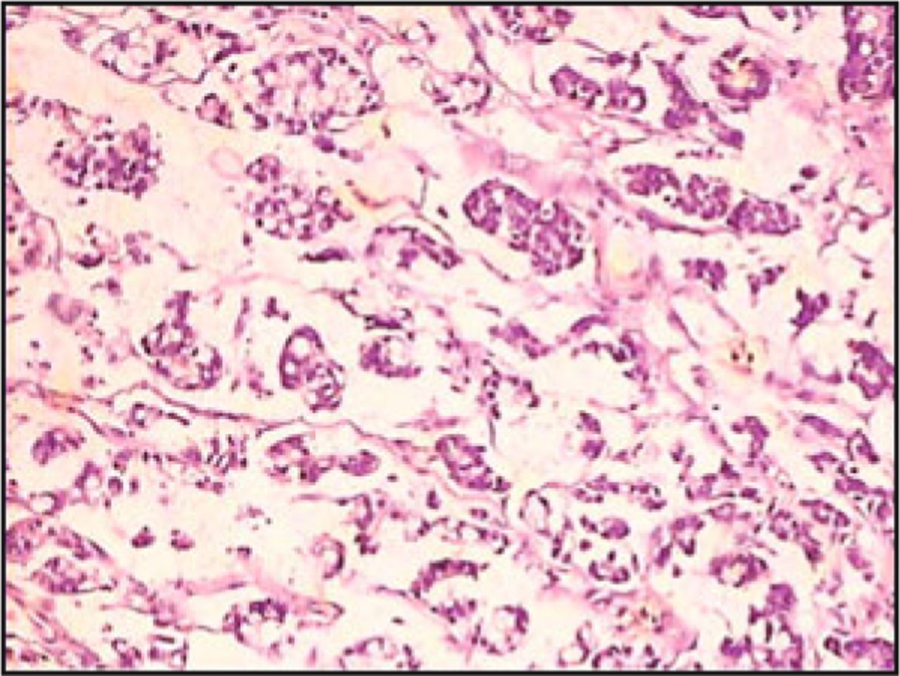
In October 2008, a 40-year-old man came to the Affiliated Hospital of Chengde Medical College for a physical examination. He indicated pain in his esophagus when swallowing food. OLYMPUS GIF-XQ260 Esophagogastroduodenoscopy was given and showed an ulcerative-type tumor about 1.5 cm in diameter on the posterior wall of his upper esophagus. Three pieces of biopsy samples were taken through esophagogastroduodenoscopy. Histological examination showed a mucosal tumor in the esophagus. Pathological diagnosis for esophageal malignancy, surgery was performed one week later. During the surgical procedure, an ulcerative-type tumor of grayish white and grayish yellow color, about 1.5 × 1 × 0.8 cm3 in size, with a tough texture was found. The specimen was inserted in 10% formaldehyde and embedded in paraffin after being removed in the surgery. After being cut into 5 μm sections the small pieces of the specimen were deparaffinized and rehydrated for hematoxylin and eosin staining. Cytokeratin, synaptophysin, and chromogranin-A (Beijing Zhongshan Biotechnology Co, LTD, The haidian district of Beijing zhongguancun 6 China) were detected in tumor tissue using 2-step immunohistochemical technique and the Dako Envision system. The positive staining of tumor cells was presented as red-brown granules, which were mainly located in cytoplasm when observed under microscope. Control sections were incubated with normal rabbit serum instead of primary antibody. Hematoxylin and eosin staining showed two types of tumor cells: carcinoid accounting for approx, 50% and adenocarcinoma accounting for the remaining 50%. Carcinoid of the esophagus presented uniform ovoid cells with cordal and small nodal patterns within the esophageal stroma (Fig. 1). The cancer tissue was shaped like rosettes when viewed under the microscope (Fig. 2). The adenocarcinoma tumor demonstrated signet ring cells and small heterogeneous glands within the esophageal stroma (Fig. 3). The immunohistochemical staining showed part of tumor synaptophysin and chromogranin-A positive in tumor cells it was diagnosed as carcinoid of the esophagus (Figs. 4 and 5). Another part of tumor negative for synaptophysin and chromogranin-A tumor cells were positive for cytokeratin (Fig. 6). At the same time adenocarcinoma of the esophagus was diagnosed, a carcinoid of the esophagus concomitant with an adenocarcinoma of the esophagus was pathologically diagnosed. Usually, carcinoma invades deep muscular layers, therefore, no metastatic lymph node was found.

Fig. 1.
  • Download figure
  • Open in new tab
  • Download powerpoint
Fig. 1.

Esophagogastroduodenoscopy showed an ulcerative type tumor about 1.5cm in diameter on the posterior wall of the upper oesophagus.

Fig. 2.
  • Download figure
  • Open in new tab
  • Download powerpoint
Fig. 2.

Carcinoid of the esophagus (about 50%) and adenocarcinoma of the oesophagus, A picture of the two components for this tumor, left for adenocarcinoma, right for carcinoid tumor (H&E stain, × 40).

Fig. 3.
  • Download figure
  • Open in new tab
  • Download powerpoint
Fig. 3.

The right for carcinoid tumor presented, carcinoid of the esophagus presented uniform ovoid cells with cordal and small nodal patterns within the esophagus stroma. Cancer tissue was looked like rosettes under microscope (H&E stain, ×100).

Fig. 4.
  • Download figure
  • Open in new tab
  • Download powerpoint
Fig. 4.

The left for adenocarcinoma tumor presented signet ring cells and small heterogeneous gland within the esophagus stroma (H&E stain, ×200).

Fig. 5.
  • Download figure
  • Open in new tab
  • Download powerpoint
Fig. 5.

The right for carcinoid tumor cells showed synaptophysin positive (IHC stain, × 200).

Fig. 6.
  • Download figure
  • Open in new tab
  • Download powerpoint
Fig. 6.

The right for carcinoid tumor showed chromogranin-A positive in tumor cells (IHC stain, ×200).

Fig. 7.
  • Download figure
  • Open in new tab
  • Download powerpoint
Fig. 7.

The left for adenocarcinoma showed cytokeratin positive in tumor cells (IHC stain, × 200).

Discussion

Carcinoid of the esophagus concomitant with adenocarcinoma of the esophagus is clinically rare. To the best of current knowledge, it has not been reported in the literature. The pathogenesis of carcinoid of the esophagus concomitant with adenocarcinoma of the esophagus is unclear.

Carcinoids of the esophagus in different locations on digestive tract may have different clinical features, including esophageal pain, vomiting, and having a choking feeling[2-5]. Carcinoid tumor associated with vascular malformation may cause esophageal bleeding[6,7]. Carcinoids in the digestive tract are classified into 3 subtypes, all of which originate from digestive tract enterochromaffin-like (ECL) cells in the digestive tract mucosa. It is well known that digestive tract ECL cells in the digestive tract mucosa synthesize and secrete 5-hydroxytryptamine (5-HT)[8-11], because the ECL cells tend to become hyperplastic. This causes 5-HT level in the blood to significantly increase, and the carcinoid symptoms are caused by excessive 5-HT level in blood production. Carcinoid syndrome usually includes flushing, diarrhea, but most digestive tract carcinoids are non-functioning neoplasms, and only about 2.4% of patients with digestive tract carcinoids present with carcinoid syndrome. This patient had never experienced any digestive tract symptoms nor carcinoid syndrome. The diagnosis of carcinoid of the esophagus concomitant with adenocarcinoma of the esophagus depended on the features of endoscopic examination, B-ultrasound, computed tomography, endoscopic retrograde cholangiopancreatography (ERCP), endoscopic ultrasonography and pathologic examination, especially immunohistochemical examination for markers, such as synaptophysin, cytokeratin, neuron-specific enolase (NSE), chromogranin-A, epithelial membrane antigen and/or neurosecretory granules, which are observed through electronic microscope.

Carcinoids of the esophagus are preferentially diagnosed as tumors based on the findings of cytokeratin, synaptophysin, chromogranin-A, and positive neurosecretory granules in immunohistochemical staining, which are observed through electronic microscope.

The treatment modality for carcinoid of the esophagus concomitant with adenocarcinoma of the esophagus is surgery[12-18] followed by chemotherapy including chemoembolization, radiotherapy and/or biotherapy. The initial treatment of functioning carcinoid of the esophagus concomitant with adenocarcinoma of the esophagus is to control the symptoms caused by excessive 5-hydroxytryptamine production.

In conclusion, the purpose of this report was to demonstrate this rare case so that other fellows in the field of oncology could learn something from it.

Conflict of interest statement

No potential conflicts of interest were disclosed.

  • Received July 11, 2010.
  • Accepted August 18, 2010.
  • Copyright © 2010 by Tianjin Medical University Cancer Institute & Hospital and Springer

References

  1. ↵
    1. Yoon JH,
    2. Lee OY,
    3. Cho SC, et al.
    A case of small duodenal carcinoid tumor with liver metastasis. Korean J Gastroenterol 2009; 53: 246-250.
    OpenUrlPubMed
  2. ↵
    1. Liang JW,
    2. Zhou ZX,
    3. Liu Q, et al.
    Clinicopathological features and prognosis of different gastric carcinoid subtypes. Zhonghua Zhong Liu Za Zhi 2008; 30: 866-869 (Chinese).
    OpenUrlPubMed
    1. Borch K,
    2. Ahrén B,
    3. Ahlman H, et al.
    Gastric carcinoids: biologic behavior and prognosis after differentiated treatment in relation to type. AnnSurg 2005; 242: 64-73.
    OpenUrlCrossRefPubMed
    1. Hoshino M,
    2. Omura N,
    3. Yano F, et al.
    Usefulness of laparoscope-assisted antrectomy for gastric carcinoids with hypergastrinemia. Hepatogastroenterology 2010; 57: 379-382.
    OpenUrlPubMed
  3. ↵
    1. Ozao-Choy J,
    2. Buch K,
    3. Strauchen JA, et al.
    Laparoscopic antrectomy for the treatment of type I gastric carcinoid tumors. J Surg Res 2010; 162: 22-25.
    OpenUrlCrossRefPubMed
  4. ↵
    1. Roncoroni L,
    2. Costi R,
    3. Canavese G, et al.
    Carcinoid tumor associated with vascular malformation as a cause of massive gastric bleeding. Am J Gastroenterol 1997; 92: 2119-2121.
    OpenUrlPubMed
  5. ↵
    1. Clemente G,
    2. Sarno G,
    3. Giordano M, et al.
    Total gastrectomy for type 1 gastric carcinoid: an unusual surgical indication? Minerva Chir 2007; 62: 421-424.
    OpenUrlPubMed
  6. ↵
    1. Li CH,
    2. Yang ZW,
    3. Yin ZR, et al.
    Relationship between atrial natriuretic peptide-immunoreactive cells and microvessels in rat gastric mucosa. Acta Pharmacologica Sinica 2006; 27: 205-211.
    OpenUrl
    1. Cooke HJ.
    Neurotransmitters in neuronal reflexes regulating intestinal secretion. AnnN Y Acad Sci 2000; 915: 77-80.
    OpenUrlPubMed
    1. Xue J,
    2. Askwith C,
    3. Javed NH, et al.
    Autonomic nervous system and secretion across the intestinal mucosal surface. Auton Neurosci 2007; 133: 55-63.
    OpenUrlCrossRefPubMed
  7. ↵
    1. Christofi FL.
    Purinergic receptors and gastrointestinal secretomotor function. Purinergic Signal 2008; 4: 213-236.
    OpenUrlCrossRefPubMed
  8. ↵
    1. Hansen MB,
    2. Witte AB.
    The role of serotonin in intestinal luminal sensing and secretion. Acta Physiol 2008; 193: 311-323.
    OpenUrl
    1. Holzer P.
    Role of visceral afferent neurons in mucosal inflammation and defense. Curr Opin Pharmacol 2007; 7: 563-569.
    OpenUrlCrossRefPubMed
    1. Kadikoylu G,
    2. Yavasoglu I,
    3. Yukselen V, et al.
    Treatment of solitary gastric carcinoid tumor by endoscopic polypectomy in a patient with pernicious anemia. World J Gastroenterol 2006; 12: 4267-4269.
    OpenUrlPubMed
    1. Somogyi A,
    2. Ruzicska E,
    3. Varga T, et al.
    Development of silent gastric carcinoid in a type 1 diabetic patient with primer hypothyreosis. Orv Hetil 2007; 148: 1667-1671.
    OpenUrlPubMed
    1. Fernández-Ruiz M,
    2. Rodríguez-Gil Y,
    3. Sáenz-López S, et al.
    Solitary gastric carcinoid tumor and pernicious anaemia: treatment by endoscopic polypectomy. Rev Esp Enferm Dig 2008; 100: 186-187.
    OpenUrlPubMed
    1. Song KY,
    2. Kim SN,
    3. Park CH.
    Laparoscopy-assisted distal gastrectomy with D2 lymph node dissection for gastric cancer: technical and oncologic aspects. Surg Endosc 2008; 22: 655-659.
    OpenUrlCrossRefPubMed
  9. ↵
    1. Kunisaki C,
    2. Makino H,
    3. Takagawa R, et al.
    Efficacy of laparoscopy-assisted distal gastrectomy for gastric cancer in the elderly. Surg Endosc 2009; 23: 377-383.
    OpenUrlPubMed
PreviousNext
Back to top

In this issue

Cancer Biology and Medicine: 7 (4)
Clinical Oncology and Cancer Research
Vol. 7, Issue 4
1 Aug 2010
  • Table of Contents
  • Index by author
Print
Download PDF
Email Article

Thank you for your interest in spreading the word on Cancer Biology & Medicine.

NOTE: We only request your email address so that the person you are recommending the page to knows that you wanted them to see it, and that it is not junk mail. We do not capture any email address.

Enter multiple addresses on separate lines or separate them with commas.
Carcinoid of the Esophagus Concomitant with Adenocarcinoma of the Esophagus: One Case Report and Literature Review
(Your Name) has sent you a message from Cancer Biology & Medicine
(Your Name) thought you would like to see the Cancer Biology & Medicine web site.
Citation Tools
Carcinoid of the Esophagus Concomitant with Adenocarcinoma of the Esophagus: One Case Report and Literature Review
Chun-hui LI, Li-hui PAN, Bei-bei XU, De-li ZHANG, Xiao-ping JIN
Clinical Oncology and Cancer Research Aug 2010, 7 (4) 271-273; DOI: 10.1007/s11805-010-0530-z

Citation Manager Formats

  • BibTeX
  • Bookends
  • EasyBib
  • EndNote (tagged)
  • EndNote 8 (xml)
  • Medlars
  • Mendeley
  • Papers
  • RefWorks Tagged
  • Ref Manager
  • RIS
  • Zotero
Share
Carcinoid of the Esophagus Concomitant with Adenocarcinoma of the Esophagus: One Case Report and Literature Review
Chun-hui LI, Li-hui PAN, Bei-bei XU, De-li ZHANG, Xiao-ping JIN
Clinical Oncology and Cancer Research Aug 2010, 7 (4) 271-273; DOI: 10.1007/s11805-010-0530-z
Twitter logo Facebook logo Mendeley logo
  • Tweet Widget
  • Facebook Like
  • Google Plus One

Jump to section

  • Article
    • Introduction
    • Case Report
    • Discussion
    • Conflict of interest statement
    • References
  • Figures & Data
  • Info & Metrics
  • References
  • PDF

Related Articles

  • No related articles found.
  • Google Scholar

Cited By...

  • No citing articles found.
  • Google Scholar

More in this TOC Section

  • Changing Paradigms in Clinical Oncology Research — Highlights from the 2011 ASCO Annual Meeting and Beyond
  • B7-H4 Expression and Increased Death Risk of Cancer Patients: A Meta-Analysis
  • Efficacy and Immune Mechanisms of Cetuximab for the Treatment of Metastatic Colorectal Cancer
Show more Research Article

Similar Articles

Keywords

  • complications
  • concomitant disease
  • esophageal neoplasms
  • adenocarcinoma

Navigate

  • Home
  • Current Issue

More Information

  • About CBM
  • About CACA
  • About TMUCIH
  • Editorial Board
  • Subscription

For Authors

  • Instructions for authors
  • Journal Policies
  • Submit a Manuscript

Journal Services

  • Email Alerts
  • Facebook
  • RSS Feeds
  • Twitter


© 2026 Cancer Biology & Medicine

Powered by HighWire