
Main menu

  • Home
  • About
    • About CBM
    • Editorial Board
    • Announcement
  • Articles
    • Ahead of print
    • Current Issue
    • Archive
    • Collections
    • Cover Story
  • For Authors
    • Instructions for Authors
    • Resources
    • Submit a Manuscript
  • For Reviewers
    • Become a Reviewer
    • Instructions for Reviewers
    • Resources
    • Outstanding Reviewer
  • Subscription
  • Alerts
    • Email Alerts
    • RSS Feeds
    • Table of Contents
  • Contact us
  • Other Publications
    • cbm

User menu

  • My alerts

Search

  • Advanced search
Cancer Biology & Medicine
  • Other Publications
    • cbm
  • My alerts
Cancer Biology & Medicine

Advanced Search


  • Home
  • About
    • About CBM
    • Editorial Board
    • Announcement
  • Articles
    • Ahead of print
    • Current Issue
    • Archive
    • Collections
    • Cover Story
  • For Authors
    • Instructions for Authors
    • Resources
    • Submit a Manuscript
  • For Reviewers
    • Become a Reviewer
    • Instructions for Reviewers
    • Resources
    • Outstanding Reviewer
  • Subscription
  • Alerts
    • Email Alerts
    • RSS Feeds
    • Table of Contents
  • Contact us
  • Follow cbm on Twitter
  • Visit cbm on Facebook
Research ArticleResearch Article

Kaposiform Hemangioendothelioma: a Case Report

Leina Sun and Zhongli Zhan
Chinese Journal of Clinical Oncology August 2006, 3 (4) 303-304;
Leina Sun
Department of Pathology, Cancer Hospital of Tiajin Medical University, Tianjin 300060, China.
  • Find this author on Google Scholar
  • Find this author on PubMed
  • Search for this author on this site
  • For correspondence: [email protected]
Zhongli Zhan
Department of Pathology, Cancer Hospital of Tiajin Medical University, Tianjin 300060, China.
  • Find this author on Google Scholar
  • Find this author on PubMed
  • Search for this author on this site
  • Article
  • Figures & Data
  • Info & Metrics
  • References
  • PDF
Loading
KEYWORDS:

keywords

  • kaposiform hemangioendothelioma
  • pathology
  • immunohisto-chemistry

Case Report

A 36-year-old man with a neoplasm in his right-leg had undergone surgical removal of this neoplasm at an out-patient clinic (OPD). The tumor size was about 1 cm× 1 cm× 0.5 cm. It was knotty and nonencapsulated. Its cross-section was kermesinus and its texture was moderate. The pathological examination confirmed a diagnosis of Kaposiform hemangioendothelioma.

Discussion

Kaposiform haemangioendothelioma is a locally aggressive, immature vascular neoplasm, characterized by a predominant Kaposi sarcoma-like fascicular spindle cell growth pattem. It also has been called Kaposi-like infantile haemangioendothelioma, or haemangioma with Kaposi sarcoma-like features. This is a rare tumor with no known racial predilection. The tumor most commonly occurs in the retroperitoneum and the skin, but it can also occur in the head and neck region, mediastinum, and deeper soft tissues of the trunk and extremities. Kaposiform haemangioendothelioma typically occurs in infancy and first decade of life, but adult cases are increasingly recognized. Retroperitoneal tumors usually present as an abdominal mass, ascites, intestinal obstruction, and jaundice. Deep soft tissue lesions produce single or multiple masses and may involve the underlying bone and rarely regional lymph nodes (variably interpreted as either local extension or local metastasis). Cutaneous lesions present as ill defined violaceous plaques. Consumption coagulopathy (Kasabach-Merritt syndrome) may complicate the larger tumors due to activation of clotting pathways within the tumor vasculature. There is no known association with HIV infection or HHV8.

Cutaneous lesions appear as ill defined, violaceous plaques. Soft tissue tumors are grayish to reddish, multi-nodular, and may coalesce and encase surrounding structures. Microscopically, the tumor grows in the form of infiltrative vague lobules separated by fibrous septa. It consists predominantly of criss-crossing spindle cell fascicles inter-spersed with capillaries. The fascicles are curved or straight, and may be compact with few interspersed spaces or more loosely arranged, containing slit-like, sieve-like or crescent-shaped vascular lumens. Nuclear atypia and mitotic activity are usually inconspicuous. Rarely, the spindle cell fascicles may blend with round “glomeruloid solid nests of polygonal/epithelioid endothelial cells which possess abundant eosinophilic cytoplasm". The interspersed capillaries are lined by flat or plump endothelial cells, and there can be tumor lobules resembling cellular haemangioma or capillary haemangioma. There are often adjacent foci resembling lymphangiomatosis. Fibrin thrombi and fragmented red cells can be found in the slit-like spaces and the capillaries. There may be haemorrhage, haemosiderin deposition and rare hyaline globules. We noted these phenomena in our case Fig.1.

Fig. 1.
  • Download figure
  • Open in new tab
  • Download powerpoint
Fig. 1.

H&E staining (200 and 400x)

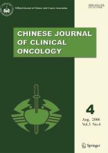
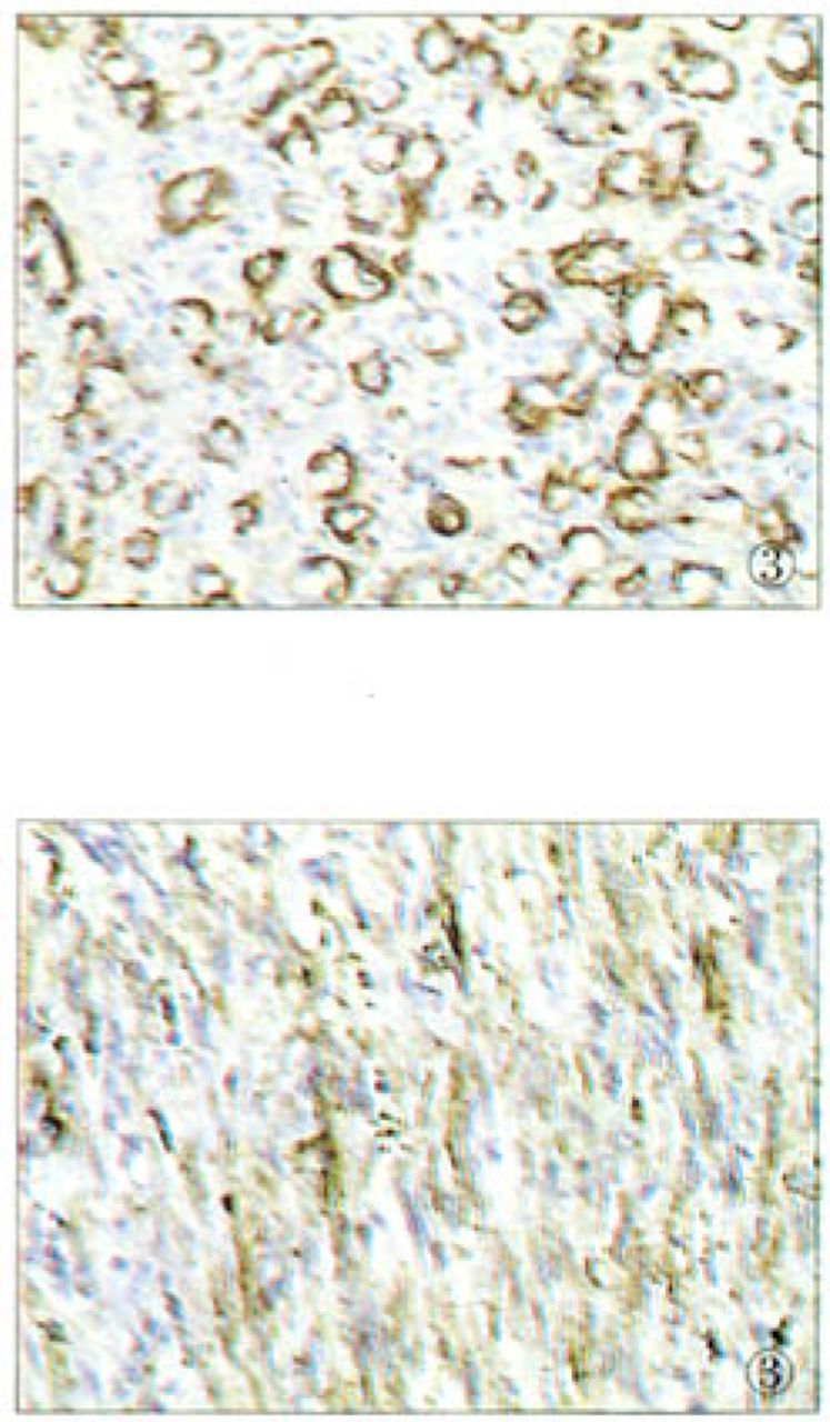
The spindle cells are usually negative for Factor VUI-related antigen, but positive for CD34 and CD 31, especially those lining vascular slits. Muscle-specific actin highlights variable numbers of spindle cells, suggesting the presence of pericytes in at least some areas. In this case, positive expression of CD34, SMA can be seen, and Factor VIII was expressed obscurely Fig.2.Fig.3. Ultrastructural hallmarks of endothelial cells are poorly developed in the spindle cells and represented by poorly formed lumens and discontinuous basal lamina. Weibel-Palade bodies may be totally absent.

Fig. 2.
  • Download figure
  • Open in new tab
  • Download powerpoint
Fig. 2.

Positive immunostaining of CD34 of the tumor cells (200x).

Fig. 3.
  • Download figure
  • Open in new tab
  • Download powerpoint
Fig. 3.

Positive immunostaining of (SMA) of the tumor cells (200* ).

Kaposiform haemangioendothelioma shows no tendency for spontaneous regression. The prognosis varies with the site and size of the lesion. Outlook is poor for large tumors occurring in infancy complicated by Kasabach-Merritt syndrome, especially when occurring in intraabdominal sites. Lesions in the somatic soft tissue are curable by complete excision, and recurrence appears to be rare.

  • Received August 3, 2006.
  • Accepted August 22, 2006.
  • Copyright © 2006 by Tianjin Medical University Cancer Institute & Hospital and Springer

References

    1. Zukerberg LR,
    2. Nickoloff BJ,
    3. Weiss SW
    . Kaposiform hemangioendothelioma of infancy and childhood. An aggressive neoplasm associated with Kasabach-Merritt syndrome and lymphangiomatosis. Am J Surg Pathol. 1993;17321.
    1. Lyons LL,
    2. North PE,
    3. Mac-Moune Lai F, et al
    . Kaposiform hemangioendothelioma: a study of 33 cases emphasizing its pathologic, immunophenotypic, and biologic uniqueness from juvenile hemangioma. Am J Surg Pathol. 2004;28:559–568.
    OpenUrlCrossRefPubMed
    1. Mentzel T,
    2. Mazzoleni G,
    3. Dei Tos AP, et al
    . Kaposiform hemangioendothelioma in adults. Clinicopathologic and immunohistochemical analysis of three cases. Am J Clin Pathol. 1997;108:450–455.
    OpenUrlCrossRefPubMed
    1. Tang HF,
    2. Zhou YY,
    3. Gu WZ
    . The clinical feature of Kaposiform hemangioendothelioma. Chin Sur J (Chinese). 2004;42:1132–1135.
    OpenUrl
    1. Cheuk W,
    2. Wong KO,
    3. Wong CS, et al
    . Immunostaining for human herpesvirus 8 latent nuclear antigen-1 helps distinguish Kaposi sarcoma from its mimickers. Am J Clin Pathol. 2004;121:335–342.
    OpenUrlCrossRefPubMed
PreviousNext
Back to top

In this issue

Cancer Biology and Medicine: 3 (4)
Chinese Journal of Clinical Oncology
Vol. 3, Issue 4
1 Aug 2006
  • Table of Contents
  • Index by author
Print
Download PDF
Email Article

Thank you for your interest in spreading the word on Cancer Biology & Medicine.

NOTE: We only request your email address so that the person you are recommending the page to knows that you wanted them to see it, and that it is not junk mail. We do not capture any email address.

Enter multiple addresses on separate lines or separate them with commas.
Kaposiform Hemangioendothelioma: a Case Report
(Your Name) has sent you a message from Cancer Biology & Medicine
(Your Name) thought you would like to see the Cancer Biology & Medicine web site.
Citation Tools
Kaposiform Hemangioendothelioma: a Case Report
Leina Sun, Zhongli Zhan
Chinese Journal of Clinical Oncology Aug 2006, 3 (4) 303-304;

Citation Manager Formats

  • BibTeX
  • Bookends
  • EasyBib
  • EndNote (tagged)
  • EndNote 8 (xml)
  • Medlars
  • Mendeley
  • Papers
  • RefWorks Tagged
  • Ref Manager
  • RIS
  • Zotero
Share
Kaposiform Hemangioendothelioma: a Case Report
Leina Sun, Zhongli Zhan
Chinese Journal of Clinical Oncology Aug 2006, 3 (4) 303-304;
Twitter logo Facebook logo Mendeley logo
  • Tweet Widget
  • Facebook Like
  • Google Plus One

Jump to section

  • Article
    • Case Report
    • Discussion
    • References
  • Figures & Data
  • Info & Metrics
  • References
  • PDF

Related Articles

  • No related articles found.
  • Google Scholar

Cited By...

  • No citing articles found.
  • Google Scholar

More in this TOC Section

  • Intussusception Induced by Transverse Colon Lipoma in a Young Male Patient—One Case Report
  • Changing Paradigms in Clinical Oncology Research — Highlights from the 2011 ASCO Annual Meeting and Beyond
  • B7-H4 Expression and Increased Death Risk of Cancer Patients: A Meta-Analysis
Show more Research Article

Similar Articles

Keywords

  • kaposiform hemangioendothelioma
  • pathology
  • immunohisto-chemistry

Navigate

  • Home
  • Current Issue

More Information

  • About CBM
  • About CACA
  • About TMUCIH
  • Editorial Board
  • Subscription

For Authors

  • Instructions for authors
  • Journal Policies
  • Submit a Manuscript

Journal Services

  • Email Alerts
  • Facebook
  • RSS Feeds
  • Twitter


© 2025 Cancer Biology & Medicine

Powered by HighWire