

Abstract
OBJECTIVE Chemotherapy is an important therapy for hepatocellular carcinoma (HCC). However, it is not effective in many cases due to recurrence and metastasis even if the initial treatment produces a response. Multidrug resistance (MDR) is considered to be one of the considerable causes. The aim of this study was to reverse MDR of HepG2/ADM cells by blocking mdrl with an adenovirus vector carrying antisense mdrl in a tumor transplantated in athymic mice.
METHODS pCMVMv E was removed from the pshuttle vector. A 0.3 kb AFP promoter was inserted into the pshuttle vector and pCMV changed into pAFP. The pAFP and asmdrl PCR products were doubly digested with Kpn and Apal, the digested products were ligated by T4 ligase, the asmdrl gene was inserted into pAFP and a newly plasmid pAFP-asmdr1 was constructed. Following digestion with Pl-Scel/I-Ceu I, pAFP-asmdr1 was ligated with Adeno-× genome DNA and amplified in E.coli XL1-Blue. The HEK293 cells were transfected and virus collected. The HepG2 MDR cells (HepG2/ADM) were induced by graded resistance to ADM and were inoculated into athymic mice. After adeno-asmdrl was injected, the expression of mdrl -mRNA and the volume of the transplantated tumor and its cells were observed.
RESULTS Following injection with Adeno-asmdrl, the tumor volume in the ADM+Adeno-asmdr1 group did not increase. However the tumor volume in the PBS plus ADM group did significantly increase (P<0.05). in the tumor xenograft cells, mdrl mRNA in the xenografts was assessed by RT-PCR and was found to be reduced at 1 week and 4 weeks in the ADM+asmdr1 group, but it was stable in the ADM group. It was only 20% in the ADM+asmdr1 group compared to the ADM group at the 4th week (P<0.05). Evidence of apoptosis was observed in the tumor xenograft cells treated with Adeno-asmdrl, but there was rare or no apoptosis in the group treated with ADM and PBS.
CONCLUSION Adenovirus carrying antisense mdrl RNA can partially reverse the MDR of HepG2/ADM cells and inhibit tumor growth by downregulating mdrl mRNA resulting in tumor cell apoptosis.
keywords
Primary hepatic carcinoma (PHC) is one of the most common, highly malignant tumors throughout the entire world. Of these tumors 90% are hepatocellular carcinoma (HCC). Many methods of treatment such as surgery, chemotherapy, radiotherapy and interventional therapy have been employed, but often with poor success. [1, 2] Besides surgery, although chemotherapy is an important therapy, the results are not ideal as many patients have a recurrence and metastasis even though the initial chemotherapy appeared successful. One of the main causes of poor results is that the tumors develop multidrug resistance (MDR).[3] In recent years, it has been shown that the expression product of the mdrl gene, P-gpl70, is related to MDR. So, it may be possible to prevent MDR if the tumor phenotype can be changed and thus increase sensitivity to chemotherapeutics. The aim of this study was to attempt to alter the MDR of HcpG2/ADM cells by blocking mdrl with adenovirus vector carrying antisense mdrl in a tumor transplantated in athymic mice.
MATERIALS AND METHODS
Animals and reagents
Eighteen male Balb/c athymic mice 4 weeks of age and weighting 20±22 g were purchased from the Chinese Academy of Science, Shanghai Center of Experimental Animals. EX Taq DNA polymerase, T4 ligase, and a DNA isolation and purification kit were purchased from Promcga (USA). Restriction endonucleases and the DNA marker were from Takara Biotechnology, Dalian (China). Lipofectamin TM 2000 was obtained from Invitrogen (USA). RPMI-1640 medium and fetal calf scrum were purchased from Hyclone (USA). MTT and PBS were procured from Sigma (USA). DMEM was purchased from GIBCO (USA) and P-gp monoclonal antibody was obtained from Chemicon (USA).
Cell lines and vectors
The hepatocellular carcinoma cell line, HcpG2, was kindly denoted by Prof Weixue Tang (Department of Pathophysiology, Chongqing Medical University). Low-passage HEK 293 cells were obtained from the Institute for Cytobiology, Chinese Academy of Sciences, Shanghai. All cells were cultured in RPMl-1640 medium containing 10% fetal calf scrum at 37 C in saturated humidified air with 5% CO2. The cells were subcultured once every 3 days. An adenovirus vector Adcno-XTM expression system was purchased from Clontcch Corp (USA).
Establishment of HepG2/ADM cells with MDR
The HepG2 MDR cell line (HcpG2/ADM) was induced by graded resistance to ADM. Concentrations of 0.01 ~ 2.0 μg/ml ADM were added to the DMEM medium containing HepG2 cells until the HepG2 cells could tolerate 2 μg/ml of ADM with 90% survival. Induction of drug resistance was stopped and the cells preserved in DMEM medium containing 0.01 μg/ml ADM. These cells were preserved for future use by freezing. They maintained MDR and were named HcpG2/ADM cells. In order to observe drug tolerance of the HcpG2/ADM cells, ADM, 5-FU, MMC and MTX were employed. The MTT assay was used to measure cellular proliferative activity. Development of color (OD) was evaluated at 490 nm 72 h later. The inhibitory rate of tumor cell growth (%) = (OD in control-OD in study)/OD in control×100%
Construction of the adenoviral vector using AFP and asmdrl
To remove the p(MVII from the pshuttlc vector, PCR was used to amplify the 99-744 region of the pshuttlc vector. Specific primers were as follows: 5’-AGC CAG TAT CTG CTC CCT GCT TGT G-3’ (sense), 5’-ATG CTA GCG GTG CCA AAA CAA ACT CCC A-3’ (antisense). A Nhcl site was introduced into the antisense primer. The pshuttlc vector and the PCR products of the 99-744 region were both doubly digested with Mlul and Nhcl, and the digested products were ligated with T4 ligase. The Mlul site was 256, and the Nhcl was 921 in the pshuttlc vector. The 256-921 region of the pshuttlc vector was removed from the plasmid, and replaced by the region of 256-744. Then the 744-921 region containing the pCMV IE was removed from the pshuttlc vector. Thus, a new plasmid pCMV (-) was constructed.
Subsequently, the pCMV(-) and AFP PCR products were doubly digested with Nhcl and Apal. The 0.3 kb AFP promoter was inserted into pCMV (-), which was called pAFP. Then the pCMV II, was replaced by the AFP promoter. Both pAFP and asmdrl PCR products were doubly digested with Kpnl and Apal, and the digested products were ligated by T4 ligase. Then the asmdrl gene was inserted into the pAFP to construct the pAFP-asmdrl.
The newly constructed plasmid pAFP-asmdrl was then doubly digested with Pl-Sccl/I-Ccu I (New England Biolabs. UK), and the purified product ligated with Adcno-X genome DNA. It was then amplified in E.coli XLl-Bluc. The HEK293 cells were transfected with recombinant adenovirus which was linearized with Pad, as described in the manual. In brief”, the HEK293 cells were cultured in a 60 mm plate, and 10 μl Pad digested Adcno-X DNA was added to the culture medium when the cells were 50-70% coníluent. The cells then were transfected with Lipofectamin and incubated. A week later, the cells were lysed with three consecutive freeze-thaw cycles, and the virus was collected from the supernatant. The titer of the virus was about 1 ×10⁷ pfu/ml, which was determined by an end-point dilution assay.
Reverse transcriptase polymerase chain reaction (RT-PCR)
RT-PCR was employed to amplify the mdrl-mRNA from mouse transplantated tumor cells. A specific mdrl gene primer was obtained from the Shanghai Boya Biologic Corporation: 5’-ATG CTC GTG TTT GGA GA-3o (sense), 5’-AGC CAG AAC ATT CTT CC-3’ (antisense). RT-PCR was performed in a 50 μl volume consisting of 1 μM of each primer, 0.2 mM of each dNTP, 1 μl 5×AMV/Tfl buffer, 2 μl 25 mM Mg-SO4, 1 μl Tfl DNA polymerase, 1 μl AMV reverse transcriptase and 2 μl DNA template. The sample was heated to 48 °C for 45 min, 94 °C for 2 min for the first chain of cDNA, followed by amplification for 40 cycles at 94 °C for 45 s, 60 °C for 30 s, 68 °C for 90 s and 1 cycle at 68 °C for 7 min and 4 °C for 8 min for the second chain. Then 5 μl of each product was analyzed by 1% agarose gel (containing 0.5 μg/ml EB) electrophoresis.
Transplantated tumor model in athymic mice
Satisfactorily growing HepG2/ADM cells were digested with 0.25% trypase and washed twice with PBS, and then inoculated subcutaneously in the left armpit of the mice using l× 107/0.1ml.. After 1 week, the transplantated tumor was examined and 2 weeks later the diameter reached 5~6 mm.
Observation of reversed MDR activity by recombined adenovirus in HepG2/ADM cells
Eighteen athymic mice bearing tumors for 2 weeks were randomly used for the following 3 groups: PBS, ADM (2.0 μg/ml) and ADM+Adeno-asmdrl (ADM was 2 μg/ml, Adeno-asmdrl was l×109 pfii/ml). The mice were injected with 0.1 ml PBS, ADM and ADM+Adeno-asmdrl and the long and short diameters (A and B) of the tumors were measured each week. Four weeks later, the athymic mice were sacrificed and tumor tissues were collected. The tumor volume was calculated (A×B72)mm7[4] the tissue examined under transmission electron microscopy and RT-PCR conducted on the tissue samples.
Statistical analysis
Data were treated with SPSS 11.0 software and expressed as the mean±SD. Differences between groups were analyzed using the t-test with P<0.05 considered as statistically significant.
RESULTS
Inhibitory effects of ADM, 5-FU, MMC and MTX on HepG2/ADM cells and HepG2 cells
The concentrations of the HepG2ADM cells or HepG2 cells were 5 × 10⁴ml. ADM, 5-FU, MMC and MTX were added at the dosage of HepG2 cells: IC50×l, IC50× 10, ICso×20 and IC«×30, respectively. With the increase in drug concentrations, the inhibitory rate in the 2 groups also increased (P<0.01). In the HepG2/ADM cells, only the high concentration of ADM had an obvious inhibitory effect, but in the HepG2 cells, the inhibition was obvious at a low concentration (Table 1). Therefore the HepG2/ADM cells had features of MDR and tolerance to ADM.
The inhibitory rate of ADM, 5-FU, MMC and MTX on HepG2/ADM and HepG2 cells (%)
Reversal of MDR by recombined adenovirus in HepG2/ADM cells
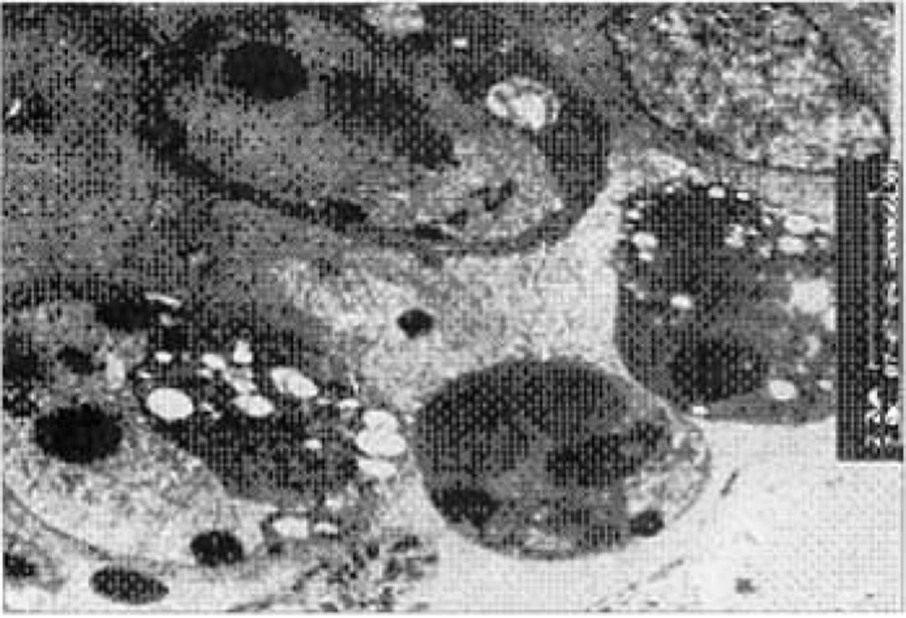
Tumor tissues were obtained from athymic mice bearing tumors for 7 days and then examined by electron microscopy. Apoptotic bodies were observed in the xenograft tumor cells treated with Adeno-asmdrl, but there was little or no apparent apoptosis in the groups treated with ADM or PBS (Fig.1).

Many apoptotic bodies were detected by transmission electron micrograph analysis in the Adeno-asmdrl group (× 8000).
The tumor volume in the ADM+Adeno-asmdrl treated group did not increase, but did obviously increase in the PBS and ADM groups (P<0.05), especially in the PBS group (Fig.2).
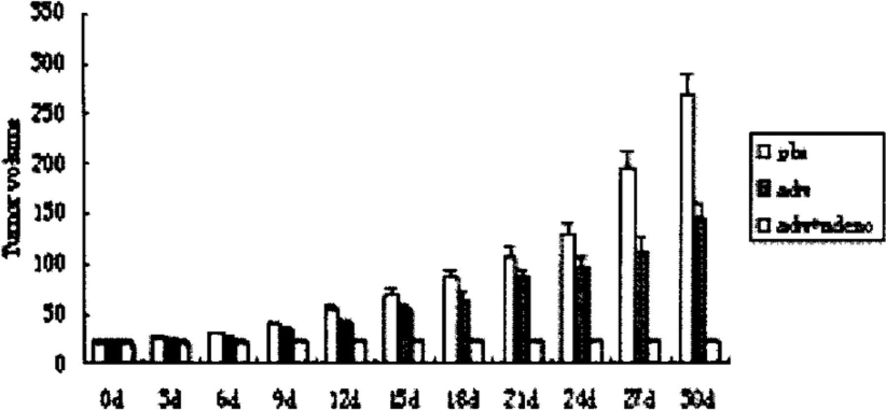
The tumor volume in the groups treated with PBS,ADM and ADM+adeno-asmdrl (P<0.05).
mdrl mRNA in xenografts examined by RT-PCR
From the electrophoresis and semi-quantitative mRNA analysis of the tumor xenografts, strips of 5 s, 18 s and 28 s were observed and the total RNA was complete. The mdrl RNA was reduced at 1 week and 4 weeks in the ADM+asmdrl group and found to be nearly unchanged in the ADM group. At 4 weeks, the mdrl RNA in the ADM+asmdrl group was only 20% compared to the ADM group (Fig.3).
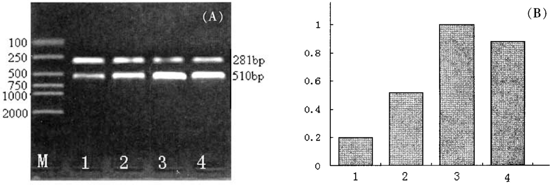
The mdrl mRNA of xenografts detected by RT-PCR (A) and by semi-quantitative mdrl mRNA analysis (B). M: marker; lane 1, ADM+asmdr1 at 4 weeks; lane 2, ADM+asmdr1 at 1 week; lane 3, ADM at 4 weeks; lane 4, ADM at 1 week.
DISCUSSION
Chemotherapy is an important mode of therapy for HCC, however, many patients do not fully respond. MDR is considered to be one of the main causes. It is known that the MDR gene family includes mdrl and mdr2, of which mdrl is the functional gene but mdr2 is non-functional.[5] Activation of the mdrl gene and overexpression of its product, P-glucoprotein (P-gp 170), play an important role in the mechanism of MDR.[6, 7] Now, many drugs are used to reverse MDR, such as the calcium channel blocker verapamil,[8] quinine,[9] naphthyridine [10] and pyronaridine,[11] but these drugs are not totally effective and have obvious side effects themselves, so their clinical use is limited.
With the development of gene transfer techniques, gene therapy is considered to be of considerable interest for HCC therapy.’[12] The distinctive expression of a unique gene is one of the important goals in tumor gene therapy. AFP is a widely accepted serologic marker for HCC. In theory, the vector carrying the AFP promoter will induce a high expression of the target gene in cells that express AFP but cause little or no expression in other cells. [13] In our study, antisense technology was employed by inserting an inverted structural gene of 200 bp from the initiation site of the coding region of mdrl. Using adeno virus as a carrier, antisense mdrl RNA was transcribed into HepG2/ ADM cells, which displayed the MDR phenotype in athymic mice. This gene transfer blocked the transcription and expression of the mdrl gene resulting in recovery of the sensitivity of the transplantated tumor to chemothcrapic agents.
Our studies showed that the volume of the transplantated tumors failed to increase when injected with a high titer of restructured adenovirus and ADM. Down-regulation of the mdrl gene transcription assessed by RT-PCR was significant compared to adminstration of only PBS or ADM. In the group receiving the restructured adenovirus plus ADM, apoptosis occurred early in the transplantated tumor. Compared with the group receiving only ADM or PBS, one can conclude that the sensitivity of the transplantated tumor to ADM had recovered as the tumor cclluar MDR was reversed by adcno-asmdrl. But this reconstructed adenovirus did not completely reverse MDR. Possible reasons for this partial reversal arc: 1) The mdrl gene is not the only gene participating in MDR, since it has been shown that a multidrug resistance-associated protein (MRP) gene [14,15] and an apoptotic regulator gene Bel-2 family [16] have an important role in MDR. Thus blocking only the mdrl gene can not totally reverse MDR. 2) There arc many concerns with regard to gene blocking technology including antisense technology, as any single therapy can not block the target gene completely.[17]”1 3) It is possible that the antisense titer was too low to block the transcription of the mdrl gene. 4) The priming efficacy of the AFP promoter was limited and could not promote down-stream transcription. 5) In this study, the treatment period was short. So, the total effect of adcno-asmdrl on MDR in the transplantated tumor and the post-effect of ADM were not observed.
Footnotes
This work was supported by the Key Program of Medical Science Foundation of Chongqing Public Health Bureau(No. [2001]01-1-018).
- Received September 28, 2005.
- Accepted December 29, 2005.
- Copyright © 2006 by Tianjin Medical University Cancer Institute & Hospital and Springer
In this issue


Jump to section
Related Articles
Cited By...
- No citing articles found.