

Abstract
OBJECTIVE To investigate apoptosis induced by Bax in hepatocellular carcinoma cells and to examine the results of 2 different routes for in vivo gene delivery.
METHODS The anti –hepatocellular carcinoma activity of the Bax gene transferred to the human hepatocellular carcinoma QGY7703 cell line was examined. In addition the Bax gene was transferred in vivo in mice via the caudal vein or hepatic artery to investigate the differences in target organ and non-target organ transfection.
RESULTS 1)The Bax gene mediated by a binary adenoviral vector system induced apoptosis in the human hepatic carcinoma QFY7703 cell line. The cell apoptotic rate in the experimental group (Bax) was 50.2±6.9% but only 32.1±9.7% in the Ad/CMV-p53 group, showing that the Bax-apoptotic rate was significantly higher than the control group. 2) LacZ expression was higher in the target organ (liver) when given through the hepatic artery than through thetail vein. In contrast, LacZ expression in the nontarget organs was higher if given through the tail vein compared to the hepatic artery.
CONCLUSION Superselective hepatic arterydelivery with Bax gene therapy is safe, specific, effective and has low toxicity. This studyprovided the basis for Bax-gene therapy via the hepatic artery in vivo.
keywords
The expression of Bax, the main member of the Bcl-2 family, can induce cellular apoptosis.It has been reported that in hepatocellular carcinoma (HCC) cells, Bax expression was reduced.[1,2] Therefore, it is an important goal to restore the apoptotic effect of Bax through a gene-transferring technique that can selectively target HCC. The technique of direct gene delivery into the hepatic artery provides a superselectively to the tumor through its nourishing artery, which is advantageous in reducing the virus distribution and expressionin non-target organs and relatively increases the designed transduction rate in the target cells.[3] In order to investigate the possibility of applying Bax gene therapy for liver cancer, the Bax gene was transferred into the QFY7703 human liver cancer cell line by a binary adenoviral vector system, with the aim of showing an apoptotic and growth-suppressing effect of the Bax gene. In addition the gene was delivered to the target organs via the tail vein or hepatic artery in rats to investigate methods of increasing expression of the gene with the goal of providing a basis for Bax gene therapy in vivo.
MATERIALS AND METHODS
Cell lines and adenoviruses
Human liver QGY7703 cancer cells were obtained from the Cancer Research Institution of Jilin, China. The cells were grown as monolayers in RPMI 1640 media supplemented with 10% heat-inactivated fetal bovine serum and antibiotics. The adenoviral vectors used in this study were the following: recombinant adenoviruses regulated by the GT minimal synthetic promoter and containing the human Bax gene cDNA (Ad/GT-Bax) or Escherichia coli β-galactosidase gene (Ad/GT-LacZ); the GV16 transactivating protein for the GT promoter under the control of the PGK promoter (Ad/PGK-GV 16); human wild-type p53 gene regulated by the immediate-early CMV promoter-enhancer (Ad/CMV-p53). These vectors were all the gifts from Dr. B.L. Fang from the University of Texas, M.D. Anderson Cancer Center, USA. All viruses were propagated in 293 cells, purified twice by ultracentrifugation in a cesium chloride gradient, and subjected to dialysis. The titer for each virus vector was determined by the absorbency of the dissociated virus at A260nm and by plaque assays. Titers for subsequent experiments were particles/ml determined by OD260nm. Particles:plaque ratios were usually between 30:1 and 100:1.
In vitro gene transfer
The QGY7703 cells were infected with Ad/GT-Bax and Ad/PGK-GV 16 at a vector ratio of 2:1 with a total multiplicity of infection (MOI) of 1000 particles. Cells treated with Ad/CMV-p53 at the same MOI were another experimental group. Cells treated with PBS or infected with Ad/GT-LacZ plus Ad/PGK-GV 16 at the same vector ratio and MOIs were used as controls.
Triggering apoptosis by induction of Bax expression
To test whether the induction of Bax expression by adenovirus-mediated gene codelivery would trigger apoptosis in the QGY7703 cells, we observed cytopathologic and morphologic changes at 48 h after treatment. In addition, the percentage of apoptotic cells was determinedby flow cytometry. Briefly, both adherent and suspended cells were harvested at 48 h after infection with viral vectors and then fixed in 70 % ethanol. Cells were stained with Hoechst 33342 and propidium iodide (PI). Apoptotic cells were quantified by flow cytometric analysisperformed at the Flow Cytometry Core Laboratory at our institution. Cell viability was determined 24, 48, and 72 h after infection by a colorimetric assay with tetrazolium dye XTT(15) using the Cell Proliferation Kit II (Roche Molecular Biochemicals, Indianapolis, IN) according to the manufacturer’s protocol. The experiments were performed at least 3 times.
Induction of Bax gene expression in vivo
To test whether Bax gene expression could be similarly induced by adenovirus-mediated gene codelivery in vivo, adult SD mice were infused via their tail vein with PBS, Ad/GT-Bax plus Ad/PGK-GV16, or Ad/GT-LacZ plus Ad/PGK-GV16 at a total vector dose of 6×l010 particles/mouse and a vector ratio of 2:1. Mice were then killed at 24 h after treatment, and liver samples taken for histopathological examination.
Induction of gene expression via the tail vein versus the hepatic artery
Selection of the route of injection that will aid in high gene expression is critical in gene therapy. Gene transduction via the hepatic artery is thought to be more efficient in liver cancer therapy than other ways. To test this hypothesis, adult SD mice were transducted with Ad/GT-LacZ at a dose of 1×10¹² particles/mouse by the hepatic artery or tail vein. After 48 h the mice were sacrificed and tissue samples taken from the liver, spleen, stomach, kidney and heart. After staining with X-Gal, 5 visual fields were chosen randomly under the light microscope. LacZ positive and negative cells were counted respectively and repeated 3 times. The transfection percentage of LacZ=LacZ positive cells/total cells×100%.
Statistical methods
Statistical evaluation was performed with the χ2 test and t test. P≤ 0.05 was considered significant.
RESULTS
Apoptosis profiles after overexpression of the Bax gene
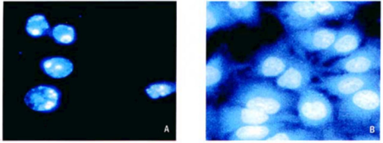
At 48 h after Bax gene transduction, changes in the cytopathology and morphology of the QGY7703 cells were observed. Over 80% of the cells treated with Ad/GT-Bax plus Ad/PGK-GV16 showed cytopatholgic signs and became rounded and detached, whereas the cells in the controlgroups remained in monolayers with normal morphology. Nuclear fragmentation, a hallmark of cell apoptosis, was detected in cells treated with Ad/GT-Bax plus Ad/PGK-GV16 (Fig.1), suggesting that Bax expression by this system did activate extensive apoptosis in this human livercancer cell line.

After transfection with Ad/GT-Bax plus Ad/PGK-GV16, QGY7703 liver cancer cells showed nuclear fragmentation, a hallmark of cell apoptosis. In the control group, the cells appeared normal. A: Ad/GT-Bax+ Ad/PGK-GV16 B: Ad/GT-Lac-Z+ Ad/PGK-GV16
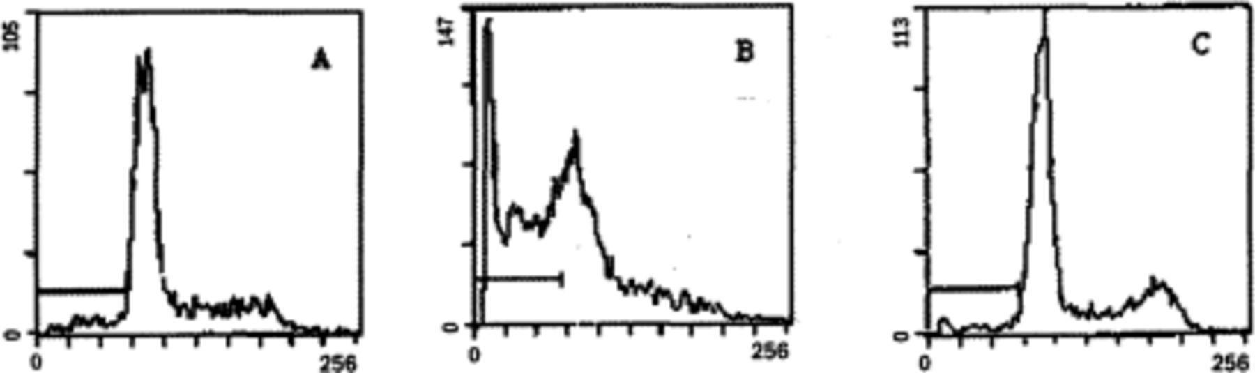
To quantify the antitumor effects of the Bax expressing vectors, the percentage of apoptotic cells in all groups was determined by flow cytometry at 48 h after treatment. While treatment with Ad/GT-LacZ plus Ad/PGK-GV16 resulted in only background levels of apoptotic cellsas that of a simulated infection, treatment with Ad/GT-Bax plus Ad/PGK-GV16 markedly increased the apoptotic cells in the QGY7703 cells (50.2%) (Fig.2) and Table 1).

Apoptotic profile. While treatment with Ad/GT-LacZ plus Ad/PGK-GV16 resulted In only background levels of apoptotic cells as that of a simulated infection, treatment with Ad/GT-Bax plus Ad/PGK-GV16 markedly increased the apoptotic cells in the QGY7703 liver cancer cells.
A: PBS; B:Ad/GT-Bax+ Ad/PGK-GV16 C: Ad/GT-Lac-Z+ Ad/PGK-GV16
To compare the antitumor effect of the Bax gene with that of the tumor suppressor p53 gene, cell viability was determined by the XTT assay at 24, 48, and 72 h after infection with Ad/CMV-p53, Ad/GT-Bax plus Ad/PGK-GV16, or Ad/GT-LacZ plus Ad/PGK-GV16.
PBS was used for a simulated control. The QGY7703 cells infected with Ad/GT-Bax plus Ad/PGK-GV16 showed massive cell death compared to Ad/CMV-p53 (Table 2), suggesting that the antitumor effect of the Bax gene is not dependent on p53 status and that the Bax gene can significantly suppress tumor growth better than p53.
Expression of the Bax gene in vivo
Expression of the Bax gene also induced typical apoptosis in normal liver cells, as revealed by nuclear fragmentation and condensation in H&E-stained liver sections (Fig.3). These results demonstrated that adenovirus-mediated gene cotransfer could produce sufficient Bax expression and induce apoptosis in vivo.

Expression of the Bax gene in normal liver cells. Expression of the Bax gene also induced typical apoptosis in normal liver cells, as revealed by nuclear fragmentation and condensation in H&E-stained liver sections.A: Ad/GT-Bax+Ad/PGK-GV16; B: Ad/GT-LacZ+Ad/PGK-GV16; C: PBS
Influence of different ways of gene transduction
To test whether there is a difference in delivering a gene through an artery or a vein, we injected Ad/GT-LacZ plus Ad/PGK-GV16 into the tail vein or the hepatic artery in adult SD mice. LacZ expression was higher in the target organ (liver) when given through the hepatic artery than through the tail vein. In contrast, LacZ expression in the nontarget organs was higher if given through the tail vein compared to the hepatic artery (Fig.4). These results suggest that transfering the gene through an artery is more efficient and has less side effects than through a vein.

A: LacZ expression was higher in liver cells through the hepatic artery. B: LacZ expression was fewer in liver cells through the tail vein.
DISCUSSION
Tumor cells have varying susceptibility to apoptosis induction. Chemotherapy or radiationtherapy may induce apoptosis in tumor cells or merely arrest the cell cycle in their normal phases, thereby opening a therapeutic window for treatment.[4]Correspondingly, insensitivity to apoptosis induction may be a major mode of resistance to antitumor therapy. The Bcl-2 family which has an important role in this process is topic of considerable research interest. Bax as the main member in this family has strong cellular toxicity, and antitumor effects and has been a concern of many researchers.[5,6]
Here we used a binary adenoviral vector system to assess the antitumor effects of the Bax gene in vitro and in vivo in the human hepatocellular carcinoma QGY7703 cell line. This vector expresses a minimal background level of Bax protein in cultured mammalian cells thus preventing apoptosis of the 293 cells, however, expression of the Bax gene can be induced substantially in vitro and in vivo by transferring it into target cells along with an adenoviral vector expressing the transactivator, fusion protein GAL4/VP16 (Ad/PGK-GV16). Because the PGKpromoter is ubiquitously active in mammalian cells, this system may also be useful for testing the antitumor activities of the Bax gene in a variety of tumor models.[7] In that research, Ad/GT-Bax+PGK-GV16 induced cell apoptosis and suppressed cell growth assessed by Hoechst 33342 staining, flow-cytometric analysis and colorimetric assay with tetrazolium dye XTT. The results we obtained demonstrated that the Bax gene can effectively induce apoptosis and suppress tumor growth both in vitro and in vivo.
The coding product of wild p53 has the capacity to monitor gene integrity, promote DNA repair and clear cells that have a cancer tendency by inducing cell apoptosis. p53 and Bax both are known to be cancer suppressor genes. But in our research, Bax and p53 had obvious differences in apoptotic sensitivity and ability to repress tumor propagation of QGY7703 cells (P <0.05). Compared to p53, the Bax gene had a low apoptotic-inducing threshold and a higher tumor-killing capacity than p53. These results suggest that the essence of Bax anti-tumor function is to induce cell apoptosis, repress cell propagation and promote cell differentiation.
As a strong pro-apoptotic gene, overexpression of the Bax gene may induce apoptosis in normal cells as well. [8] In fact, our study demonstrated that intravenous infusionof the Bax-expressing adenoviral vector induced a rapid and massive apoptosis in normal hepatocytes. The results we obtained demonstrated that expression of the Bax gene can effectively induce apoptosis and suppress tumor growth in both cancer and normal cells. This raises anissue of safety if the Bax gene is used as a therapeutic agent. Thus, targeted expression of the Bax gene is highly desirable.
An important aspect of gene therapy is to search for a safe, valid, targeting gene-transferring pathway. HCCs have abundant new blood vessels and the hepatic artery supplies over 80% of the tumor blood supply,[9] thus the anatomical basis for transferring genesvia the hepatic artery. Our results showed that under the same viral titre, the 2 different gene transferring paths varied in transferring efficiency. Compared with the tail vein pathway, LacZ distribution and expression if given via the hepatic artery is higher in the liver cells and lower in other organs. These results have important clinical significance because unresectable primary cancers in the brain, lung, pancreas, head, and neck etc. remain the major cause of morbidity and mortality in cancer patients and present most formidable problems. In liver cancers, despite the use of chemotherapy and radiation therapy in combined modality protocols, local control rates are less than 20%.[10] However, treatment of metastatic tumors by systemic gene delivery remains challenging. Our experimental results have an important significance in safety, low toxicity, targeting, validity of Bax gene therapy and provide a basis for its clinical application. Development of vector targeting and targeted transgene expression used alone or combined with other approaches may be a valuable therapeutic approach for various cancers.
ACKNOWLEDGMENTS
We thank Professor Gai Xueliang and Fan Zhimin from the Chinese-Canadian Tumor Center for statistical instruction.
Footnotes
This study was supported by the ScientifiC Research Grant from the Medical Institution of Jilin Province.
- Received April 10, 2005.
- Accepted May 12, 2005.
- Copyright © 2005 by Tianjin Medical University Cancer Institute & Hospital and Springer
In this issue


Jump to section
Related Articles
Cited By...
- No citing articles found.