

Abstract
OBJECTIVE 1) To study the efficacy of GSPs on the migration of highly metastatic mammary carcinoma cells and 2) To investigate inhibition mechanisms.
METHODS Cell migration was assessed using a 24-well transwell assay. Cells with different concentrations of GSPs were suspended (5 × 10⁵cell/mL) in RPMI 1640 media in the upper chamber, and RPMI 1640 media with 10% FBS was supplemented in the lower chamber. Then, cells were allowed to migrate for 24 h.
RESULTS GSPs inhibited the migration of 4T1 cells in a dose-dependent manner. The migration of 4T1 cells was obviously inhibited by GSPs, even at a very low concentration (5 μg/mL), and was totally inhibited when the concentration was 20 μg/mL. Also, 20 μg/mL of GSPs inhibited cell viability by only 11.4% and induced early apoptosis by only 5.6% compared with a percentage of 4.0% in control cells. GSPs suppressed the activation of PDK1, Akt and Erk1/2 in a dose-dependent manner.
CONCLUSION GSPs significantly inhibit the migration of highly metastatic mammary carcinoma 4T1 cells in vitro. This inhibition is independent of decreased cell viability or apoptosis induction. The inhibition of migration by GSPs is involved in blocking the PI3k/Akt and MAPK pathways.
keywords
Introduction
Breast cancer, one of the most common cancers worldwide, accounts for approximately 20% of female cancer mortality and is capable of metastasizing to local lymph nodes as well as to distal organs, such as bone, lung and liver[1]. Approximately 76% of all breast tumors have been categorized as invasive breast cancers. Metastasis is a complex and multi-step dispersion process by which malignant tumor cells from the primary tumor site spread to a secondary site within the body[2]. Cell migration is a critical process of invasion, allowing primary tumors to metastasize. Although several modalities, including surgery, hormone therapy and radiation therapy, are available for the treatment of breast cancer, these treatment modalities are not effective once metastasis has occurred.
Epidemiologic studies indicate that dietary habits play crucial roles in both the induction and the prevention of cancer, including breast cancer[3,4]. Therefore, efforts are needed to develop newer and effective botanicals, particularly dietary supplements, which can reduce the risk of breast cancer incidence and metastasis.
To that end, grapes, one of the most widely consumed fruits in the world, have enormous health benefits. Grape skin polyphenols (GSPs) are mainly composed of anthocyanidins and anthocyanins, which are natural pigments with antioxidant properties that also promote cancer-fighting effects[5]. Some studies have shown that GSPs inhibit the growth of mammary carcinoma cells [6]. However, their efficacy on metastasis and migration of cancer cells remain unknown.
Mouse mammary carcinoma 4T1 cells have been employed to examine the therapeutic efficacy and molecular mechanisms of chemotherapeutic agents that are relevant to humans[7-9]. The 4T1 cells are highly metastatic and poorly immunogenic and exhibit characteristics that resemble those of stage IV breast cancer in humans[10-13]. Therefore, we examined the chemoprotective efficacy of GSPs in 4T1 cells. Here, we report that in vitro treatment of 4T1 breast cancer cells with GSPs significantly inhibits their migration.
Materials and Methods
Chemicals and antibodies
The grape skin extract used in this study was obtained from JF-NATURAL Corporation (Tianjin, CN), which contained 3.1% catechin, 3.5% epicatechin, 1.8% gallic acid, 3.3% total anthocyannins, and 88.5% total phenols. The Annexin V-FITC-labeled Apoptosis Detection Kit was from BD Pharmingen (Chicago, IL). Antibodies against Akt, pAkt, PDK1, pPDK1, Erk1/2 and pErk1/2 were from Cell Signaling Technology (Beverly, MA). The respective secondary antibodies were obtained from Santa Cruz Biotechnology (Santa Cruz, CA). MTT (3-[4, 5-dimethylthia zol-2-yl]-2, 5-diphenyl tetrazolium bromide) was purchased from Beyotime (Jiangsu, CN).
MTT assay
The effect of GSPs on cell viability was determined using the MTT assay. Briefly, 2 × 10⁴ cells per well were placed in 96-well culture plates and incubated overnight. GSPs were then added to attain a final concentration ranging from 5 to 100 μg/mL, and the cells were incubated for another 48 h. At the end of the stipulated period, MTT (50 mL of 5 mg/mL) was added into each well, and the resulting formazan was then dissolved in 150 ml of dimethyl sulfoxide and the absorbance recorded at 490 nm using a Labsystems Dragon Wellscan MK-3 microplate reader.
Quantification of apoptosis by flow cytometry
The Annexin V-FITC-labeled Apoptosis Detection Kit (BD Pharmingen, Chicago, IL) was used according to the manufacturer’s instructions. Briefly, 1× 10⁵ 4T1 cells seeded in 24-well plates were treated with GSPs (10 to 50 μg/mL) for 24 h. At the end of the experiment, cells were diluted at a density of 5 × 10⁵ cells/mL in 1× supplied binding buffer and stained simultaneously with FITC-labeled Annexin V and PI. Cells were acquired using a BD FACSARIA flow cytometer. Data were analyzed with WinMdi software.
Migration assay
Migration of 4T1 cells in the presence of GSPs was assessed using 24-well Transwell permeable supports with multiporous (8-μm pore size) polycarbonate membranes (Corning, NY). Cells were suspended (5 × 10⁵ cell/mL) in RPMI 1640 media supplemented with 0.1% BSA. From this suspension, 100 Μl was plated on the upper chambers of the Transwell system with GSP concentrations of 0.1, 0.5, 1, 5, 1, 10 or 20 μg/mL respectively, and 600 μL of RPMI 1640 media supplemented with 10% FBS was placed in the lower chamber. All cells were incubated at 37°C under 5% CO2 and allowed to migrate for 24 h. At 24 h, non-migrant cells were gently swabbed off of the insert membranes, and the remaining migratory cells were then fixed and stained using hematoxylin. Migratory cells on each insert membrane were counted using a light microscope (100 × magnification). Each condition was performed in triplicate, and each migration assay was repeated three times.
Preparation of cell lysates and western blot analysis
The 4T1 cells were grown in complete DMEM medium and incubated in the absence or presence of GSPs (5, 10, 20, or 50 μg/mL) for 24 h. Thereafter, the cells were harvested, and lysates were prepared using a cell lysis buffer (Beyotime, Jiangsu, CN). The concentration of protein in the lysates was determined using the Beyotime BCA Protein Assay Kit according to the manufacturer’s protocol. For western blot analysis, an appropriate amount of lysate (30 μg protein) was subjected to SDS-PAGE using a 10% gel. Thereafter, separated proteins were transferred electrophoretically to nitrocellulose membranes. Non-specific sites on the membrane were blocked by incubation with a blocking buffer (5% non-fat dry milk in PBST) for 1 h. The membranes were then probed overnight at 4°C with the primary antibody. After washing, the membranes were incubated with the appropriate horseradish peroxidase-conjugated secondary antibody for 1 h and washed. The expression of the protein was determined using the ECL detection system and autoradiography with X-OMAT BT film (Kodak).
Results
GSPs decrease the viability of 4T1 cells
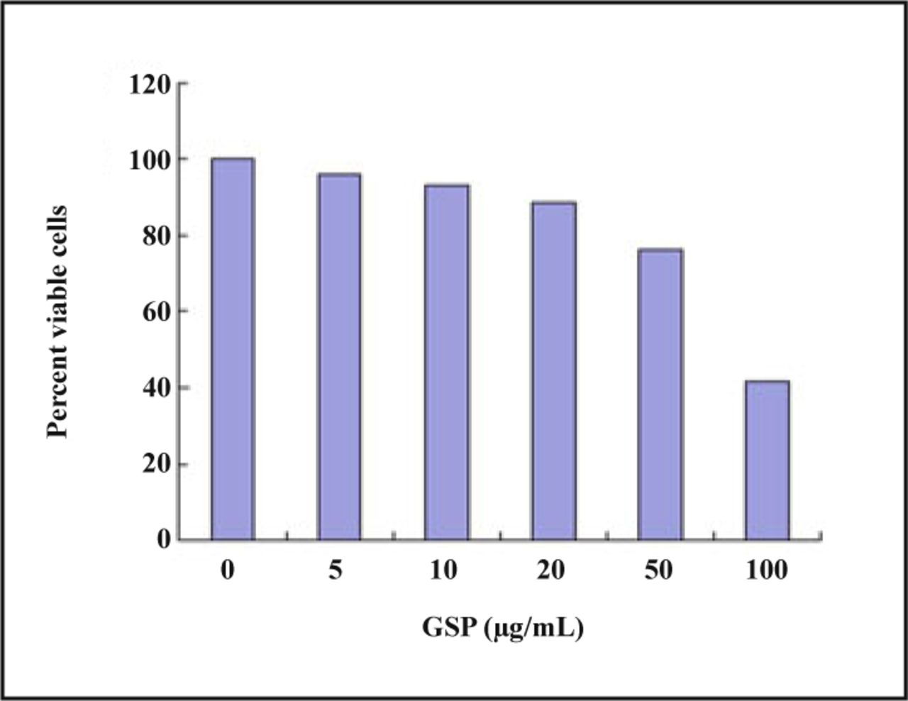
The effect of GSPs on the cell viability of 4T1 cells was confirmed using the MTT assay. As shown in Fig.1, treatment of 4T1 cells with GSPs (5-100 μg/mL) resulted in an evident reduction in cell viability in a concentration-dependent manner. The IC50 (half maximal inhibitory concentration) of GSPs is 72.408 μg/mL. These results indicate the cytotoxic activity of GSPs against highly metastatic breast cancer cells. Since our interest was to examine whether GSPs have the ability to oppose the metastatic potential of tumor cells, we selected mouse 4T1 cells, which are highly metastatic in nature, for further studies.

GSPs inhibited the viability of 4T1 cells in a dose-dependent manner. 4T1 cells were treated with GSPs at concentration ranging from 5 to 100 μg/mL for 48 h. The MTT assay was used to detect cell viability.
GSPs induce apoptosis in 4T1 cells
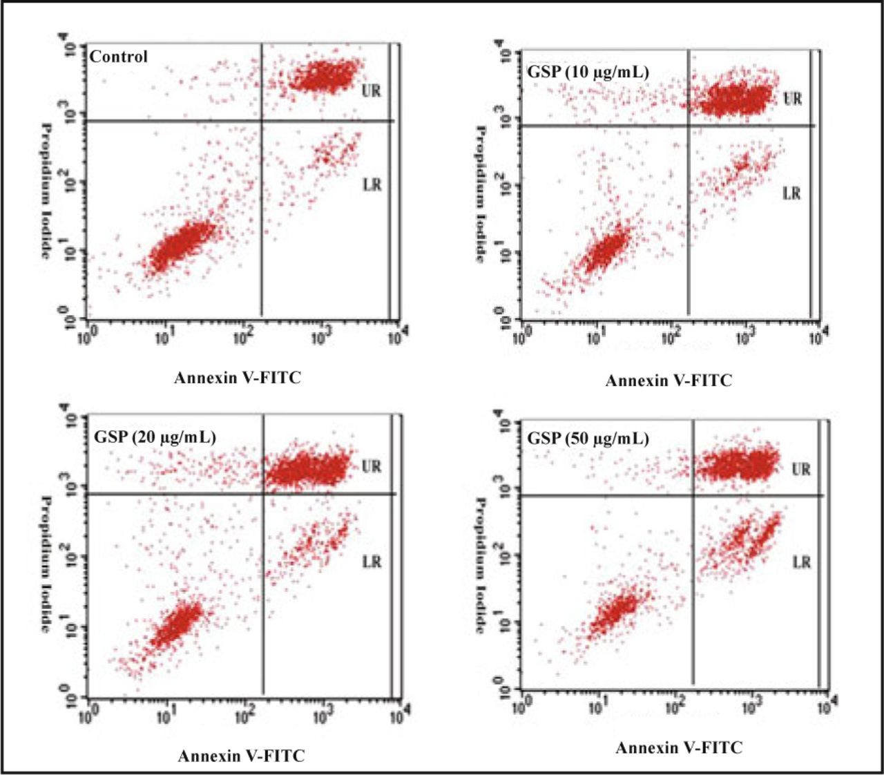
The early apoptotic cells are shown in the lower right (LR) quadrant of the histograms (Fig.2), whereas late apoptotic cells are shown in the upper right (UR) quadrant. We found that the treatment of 4T1 cells with GSPs at concentrations of 10, 20 and 50 μg/mL for 24 h resulted in an increased percent of early apoptotic cells (LR) from 5.04% to 10.76%, compared with a percentage of 4.04% in control cells that were not treated with GSPs. Similarly, treatment with GSPs did not result in an evident dose-dependent increase in the numbers of late apoptotic cells (UR). Even though the percentage of all apoptotic cells (UR + LR) was higher (42.6% for 4T1 cells treated with 50 mg/ml GSPs for 24 h) than for 4T1 cells not treated with GSPs (27.6%), GSPs at concentrations up to 50 μg/mL did not significantly induce apoptosis in 4T1 cells.

GSPs induce apoptosis of 4T1 cells after 24 h of treatment in vitro. The Annexin V-FITC-labeled Apoptosis Detection Kit was used to analyze the number of apoptotic cells by flow cytometry. The lower right (LR) quadrant of the histograms indicates the percentage of early apoptotic cells, and the upper right (UR) quadrant indicates the percentage of late apoptotic cells. For control, LR = 4.04% and UR = 23.51%; for GSP-treated cells (10 μg/mL), LR=5.04% and UR = 31.84%; for GSP-treated cells (20 μg/mL), LR=5.64% and UR = 33.80%; for GSP-treated cells (50 μg/mL), LR = 10.76% and UR = 34.94%.
GSPs suppress the migration of 4T1 cells through inhibition of the PI3K/Akt and MAPK/ERK1/2 pathways
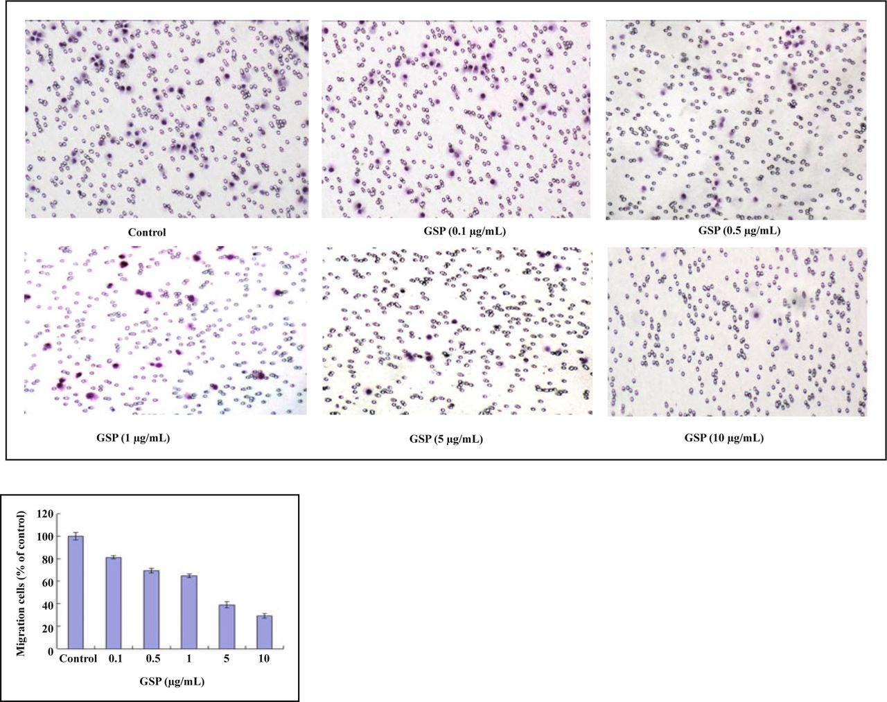
Cell migration is the critical process of invasion, allowing primary tumors to metastasize. Thus, the efficacy of GSPs on the migration of 4T1 mouse carcinoma cells was examined. The treatment of 4T1 cells with increasing concentrations of GSPs led to a significant concentration-dependent decrease in cell migration using the Boyden chamber assay (Fig.3). GSPs exhibited evident inhibition of migration, even at very low concentrations (5 μg/mL). The migration of 4T1 cells was totally inhibited when treated with GSPs at a concentration of 20 μg/mL (data not shown).
GSPs inhibit the migration of 4T1 cells in a dose-dependent manner. The cells were cultured in the presence of various concentrations of GSPs for 24 h in a Boyden chamber. Photographs of cell migration through the polycarbonate membrane stained by hematoxylin (A) and quantitative analysis of the Boyden chamber assay (B) are shown.
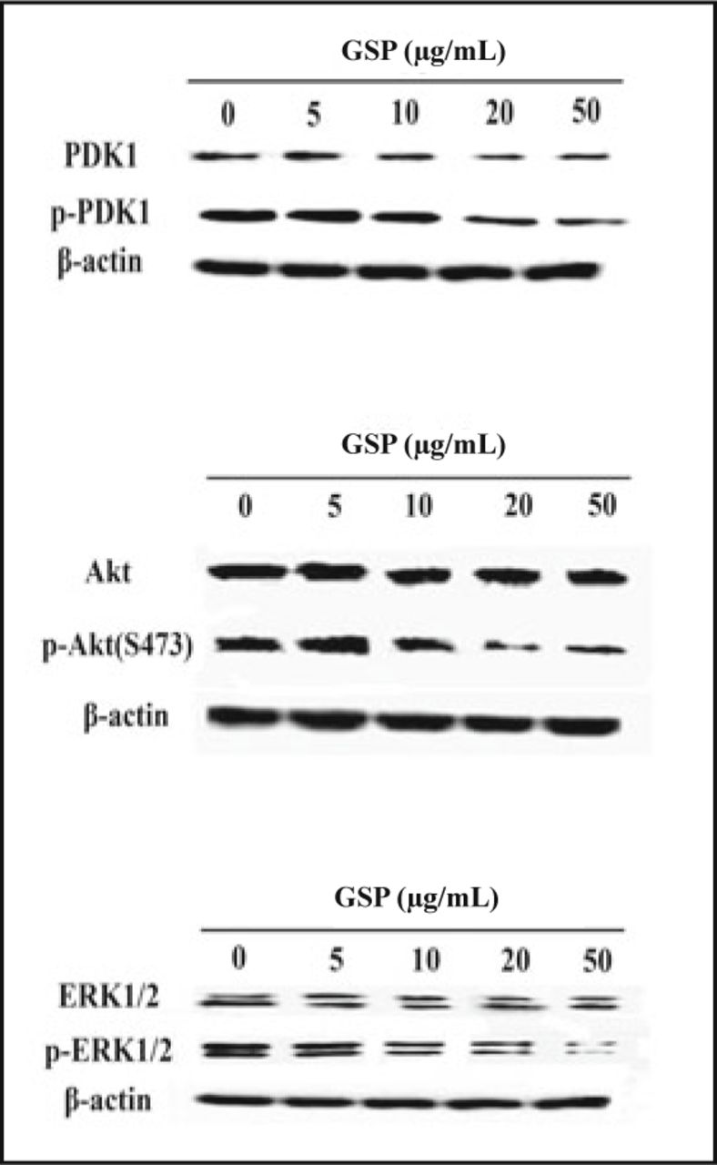
It remains to be determined which pathways are inhibited by GSPs. In order to address the molecular mechanisms by which GSPs inhibit migration of 4T1 cells, we investigated the effects of GSPs on the phosphorylation of Akt/PDK1, which is an upstream regulator of Akt in the PI3K/Akt pathway, and ERk1/2. We demonstrated that GSPs suppress activation of PDK1, Akt and ERK1/2 (Fig.4). The total protein levels of PDK1, Akt and ERK1/2 remained unchanged with these treatments. These results indicate that GSPs block the PI3K/Akt and MAPK pathway and contribute to the inhibition of cellular migration.

GSPs suppress the activation of PDK1, Akt and ERK1/2 in a dose-dependent manner. The 4T1 cells were grown in complete DMEM medium and incubated in the absence or presence of GSPs (0.1, 0.5, 1, 5 and 10 μg/mL) for 24 h. Western blot assays were used to examine total expression and phosphorylation of PDK1, Akt and ERK1/2 in 4T1 cells.
Discussion
The presence of metastasis is the main cause of morbidity and mortality in millions of patients with breast cancer. Metastasis is a complex, multi-step process involving cell adhesion, invasion and migration. Hence, interruption of one or more of these steps is one approach for anti-metastatic therapy. The present study is the first to demonstrate that grape skin polyphenols (GSPs) can significantly inhibit the migration of 4T1 mouse breast carcinoma cells.
Grape skin contains different phenolic compounds, including anthocyanins, flavonol derivatives (quercetin and kaempferol), catechin, epicatechin, resveratrol and chlorogenic acid[14]. Some studies indicate that GSPs decrease the migration of cancer cells, such as human U-87 glioblastoma and human DAOY medulloblastoma cells[15]. To date, there is no direct evidence to show that GSPs exert such an effect on the development of breast cancer metastases. This investigation attempted to address the role of GSPs in blocking the migration of highly metastatic mammary carcinoma cells, and the mechanisms by which it may exert its anti-metastatic effects.
GSPs exhibited an evident ability to inhibit the migration of 4T1 cells even at very low concentration (5 μg/mL) (Fig.3). The migration of 4T1 cells was totally inhibited when treated by GSPs at a concentration of 20 μg/mL (data not shown); this concentration is far below that of the IC50 (72.408 μg/mL). Also, 20 μg/mL of GSPs inhibit cell viability by only 11.4% (Fig.1), and induce early apoptosis only in 5.6% of cells compared with a percentage of 4.0% in control cells (Fig.2). These results indicate that the ability of GSPs to inhibit the migration of 4T1 cells was not a result of inhibited cell viability of apoptosis induction.
The PI3K/Akt and MAPK signaling pathways are crucial for many aspects of cell growth and survival[16]. In breast cancer, the PI3K/Akt pathway contributes to metastatic competence and therapy resistance[17]. Therefore, the development of phytochemicals for the treatment of breast cancers that can block PI3K/Akt and MAPK signaling will be highly beneficial. Our results reveal that GSPs suppress the activation of PDK1, Akt and ERK1/2 in a dose-dependent manner. The present study demonstrates that the migration inhibition of GSPs on 4T1 cells is a result of blocking the PI3k/Akt and MAPK pathways.
Although these findings are not expected to be directly representative of physiological effects because neither the bioavailability nor the systemic metabolism of GSPs was taken into account, they suggest that GSPs may contribute to the prevention of the migration of highly metastatic mammary carcinoma cells. Our results may be relevant for breast cancer therapy.
Conflict of interest statement
No potential conflicts of interest were disclosed.
- Received February 18, 2011.
- Accepted March 14, 2011.
- Copyright © 2011 by Tianjin Medical University Cancer Institute & Hospital and Springer
In this issue


Jump to section
Related Articles
Cited By...
- No citing articles found.