

Main menu

  • Home
  • About
    • About CBM
    • Editorial Board
    • Announcement
  • Articles
    • Ahead of print
    • Current Issue
    • Archive
    • Collections
    • Cover Story
  • For Authors
    • Instructions for Authors
    • Resources
    • Submit a Manuscript
  • For Reviewers
    • Become a Reviewer
    • Instructions for Reviewers
    • Resources
    • Outstanding Reviewer
  • Subscription
  • Alerts
    • Email Alerts
    • RSS Feeds
    • Table of Contents
  • Contact us
  • Other Publications
    • cbm

User menu

  • My alerts

Search

  • Advanced search
Cancer Biology & Medicine
  • Other Publications
    • cbm
  • My alerts
Cancer Biology & Medicine

Advanced Search


  • Home
  • About
    • About CBM
    • Editorial Board
    • Announcement
  • Articles
    • Ahead of print
    • Current Issue
    • Archive
    • Collections
    • Cover Story
  • For Authors
    • Instructions for Authors
    • Resources
    • Submit a Manuscript
  • For Reviewers
    • Become a Reviewer
    • Instructions for Reviewers
    • Resources
    • Outstanding Reviewer
  • Subscription
  • Alerts
    • Email Alerts
    • RSS Feeds
    • Table of Contents
  • Contact us
  • Follow cbm on Twitter
  • Visit cbm on Facebook
Research ArticleOriginal Article

PADI3 induces cell cycle arrest via the Sirt2/AKT/p21 pathway and acts as a tumor suppressor gene in colon cancer

Xiaotian Chang, Zhengbin Chai, Jiaorui Zou, Hongxing Wang, Yao Wang, Yabing Zheng, Hui Wu and Chunyan Liu
Cancer Biology & Medicine November 2019, 16 (4) 729-742; DOI: https://doi.org/10.20892/j.issn.2095-3941.2019.0065
Xiaotian Chang
1Medical Research Center, The First Affiliated Hospital of Shandong First Medical University, Jinan 250014, China
2Medical Research Center of the Hospital Affiliated to Qingdao University, Qingdao 266000, China
  • Find this author on Google Scholar
  • Find this author on PubMed
  • Search for this author on this site
Zhengbin Chai
3Department of Laboratory Medicine, Jinan Infectious Disease Hospital, Shandong University, Jinan 250021, China
  • Find this author on Google Scholar
  • Find this author on PubMed
  • Search for this author on this site
Jiaorui Zou
1Medical Research Center, The First Affiliated Hospital of Shandong First Medical University, Jinan 250014, China
  • Find this author on Google Scholar
  • Find this author on PubMed
  • Search for this author on this site
Hongxing Wang
1Medical Research Center, The First Affiliated Hospital of Shandong First Medical University, Jinan 250014, China
  • Find this author on Google Scholar
  • Find this author on PubMed
  • Search for this author on this site
Yao Wang
1Medical Research Center, The First Affiliated Hospital of Shandong First Medical University, Jinan 250014, China
  • Find this author on Google Scholar
  • Find this author on PubMed
  • Search for this author on this site
Yabing Zheng
1Medical Research Center, The First Affiliated Hospital of Shandong First Medical University, Jinan 250014, China
  • Find this author on Google Scholar
  • Find this author on PubMed
  • Search for this author on this site
Hui Wu
1Medical Research Center, The First Affiliated Hospital of Shandong First Medical University, Jinan 250014, China
  • Find this author on Google Scholar
  • Find this author on PubMed
  • Search for this author on this site
Chunyan Liu
1Medical Research Center, The First Affiliated Hospital of Shandong First Medical University, Jinan 250014, China
  • Find this author on Google Scholar
  • Find this author on PubMed
  • Search for this author on this site
  • For correspondence: liuchunyan2018{at}126.com
  • Article
  • Figures & Data
  • Info & Metrics
  • References
  • PDF
Loading

Article Figures & Data

Figures

  • Tables
  • 1
    • Download figure
    • Open in new tab
    • Download powerpoint
    1

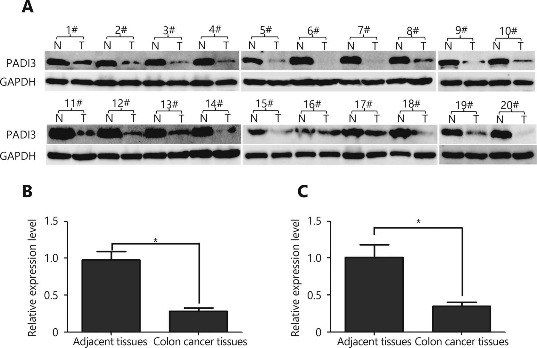
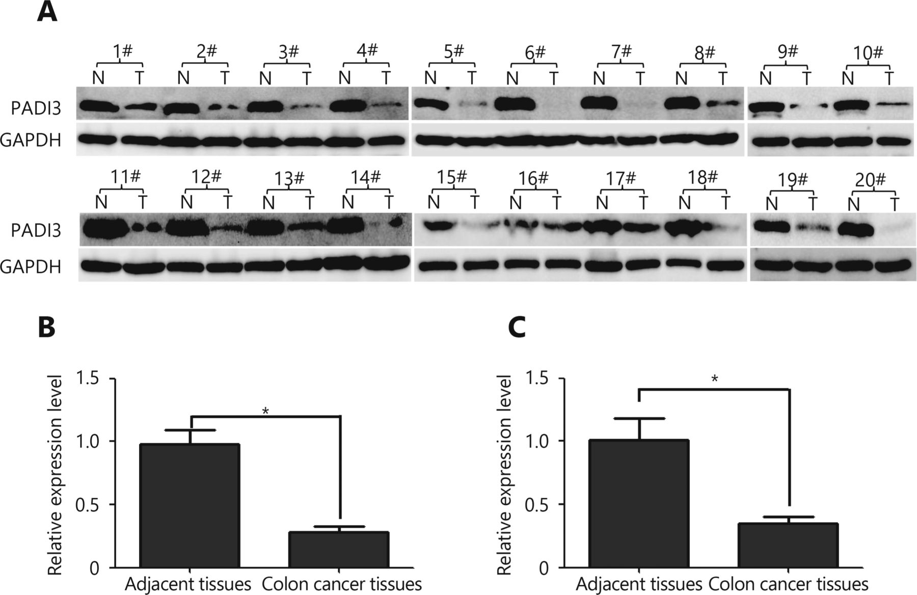
    PADI3 expression profile determination using Western blot analysis and qRT-PCR. (A) Western blot analysis was used to measure the expression level of PADI3 in colon cancer tissue samples and corresponding adjacent tissue samples at the translational level. These paired tissue samples were obtained from 20 different patients; GAPDH was selected as the internal control. (B) Statistical analysis of Western blot. (C) qRT-PCR was used to measure the expression level of PADI3 in the colon cancer tissue samples and corresponding adjacent tissue samples at the transcriptional level. T: tumor tissues; N: corresponding adjacent tissues. * indicates P < 0.05, and ** indicates P < 0.01 for three independent experiments analyzed by Student’s t-test.

  • 2
    • Download figure
    • Open in new tab
    • Download powerpoint
    2

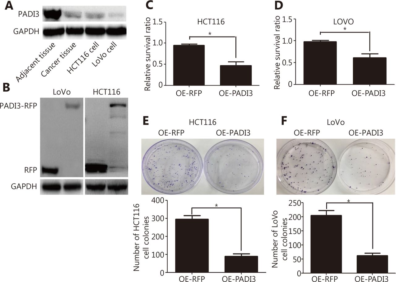
    Effects of PADI3 on HCT116 and LoVo cells. RFP-expressing cells were used as controls. (A) Weak PADI3 expression was detected in HCT116 and LoVo cells. (B) Western blot analysis was used to measure PADI3 and RFP expression in both LoVo and HCT116 cells. (C, D) A CCK-8 assay measured the proliferation of HCT116 and LoVo cells. (E) The colony formation ability of HCT116 cells was measured and statistically analyzed using a colony formation assay following 14 days of culture. (F) The colony formation ability of LoVo cells was measured and statistically analyzed using a colony formation assay following 14 days of culture. * indicates P < 0.05 for three independent experiments analyzed by Student’s t-test.

  • 3
    • Download figure
    • Open in new tab
    • Download powerpoint
    3

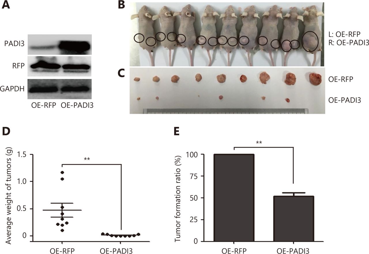
    Effects of PADI3 in vivo. (A) Western blot analysis was used to measure PADI3 expression. (B) PADI3-expressing HCT116 cells were injected into the left dorsal flank of BALB/c nude mice, and RFP-expressing HCT116 cells were injected into the right dorsal flank. (C) Tumors were dissected 42 days after cell injection. (D) Tumor weight was measured and statistically analyzed. (E) The tumor formation ratio was statistically analyzed. * indicates P < 0.05.

  • 4
    • Download figure
    • Open in new tab
    • Download powerpoint
    4

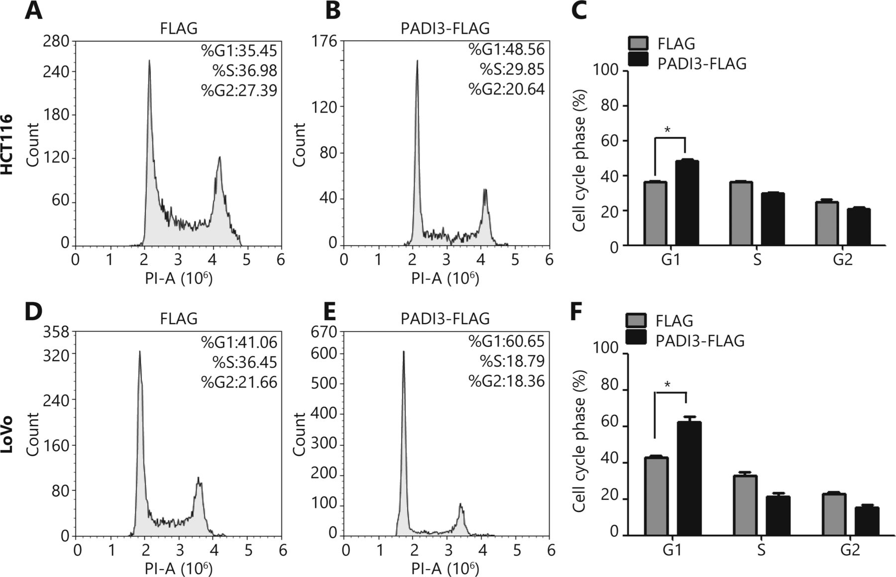
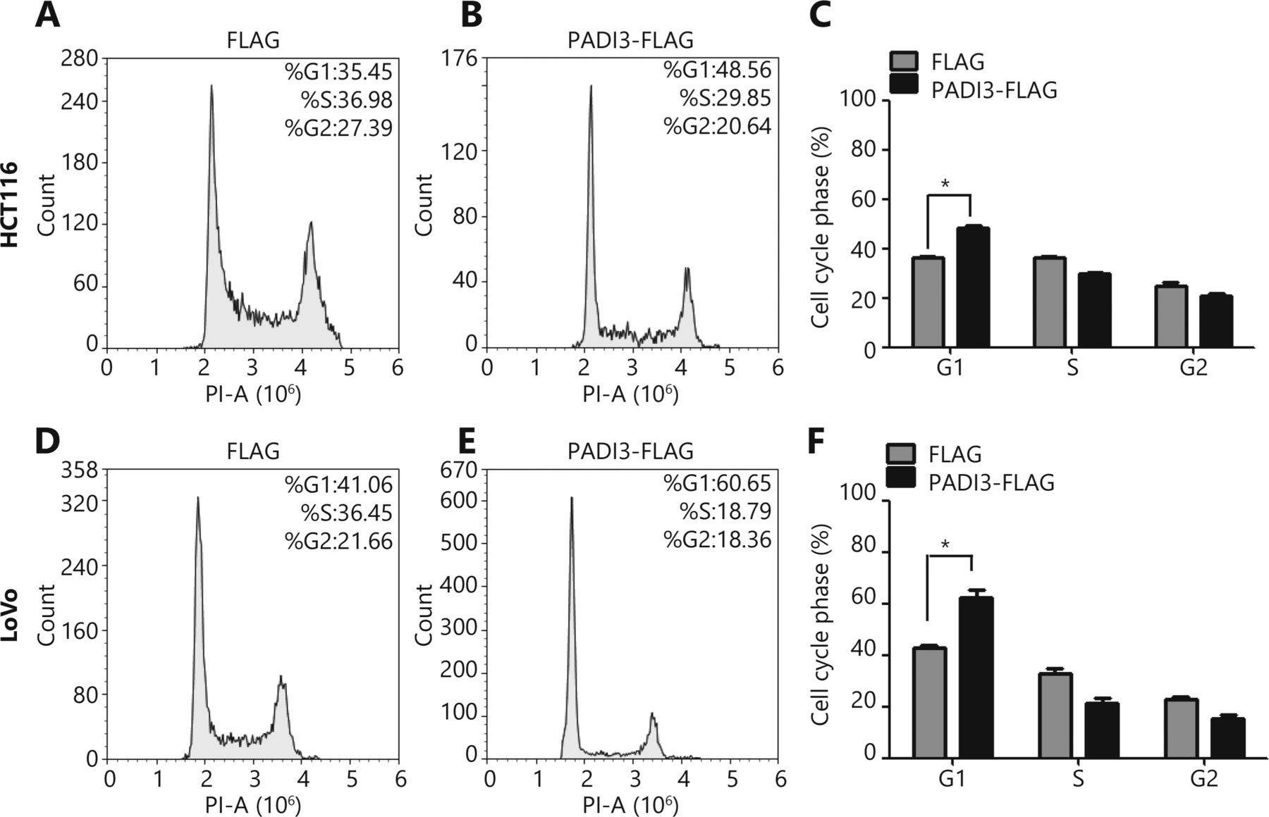
    Analysis of the roles of PADI3 in the cell cycle using FCM. (A) FLAG-expressing HCT116 cells were used as the negative control group. (B) PADI3-FLAG-expressing HCT116 cells were used as the experimental group. (C) Statistical analysis of A and B. (D) FLAG-expressing LoVo cells were used as the negative control group. (E) PADI3-FLAG-expressing LoVo cells were used as the experimental group. (F) Statistical analysis of D and E.

  • 5
    • Download figure
    • Open in new tab
    • Download powerpoint
    5

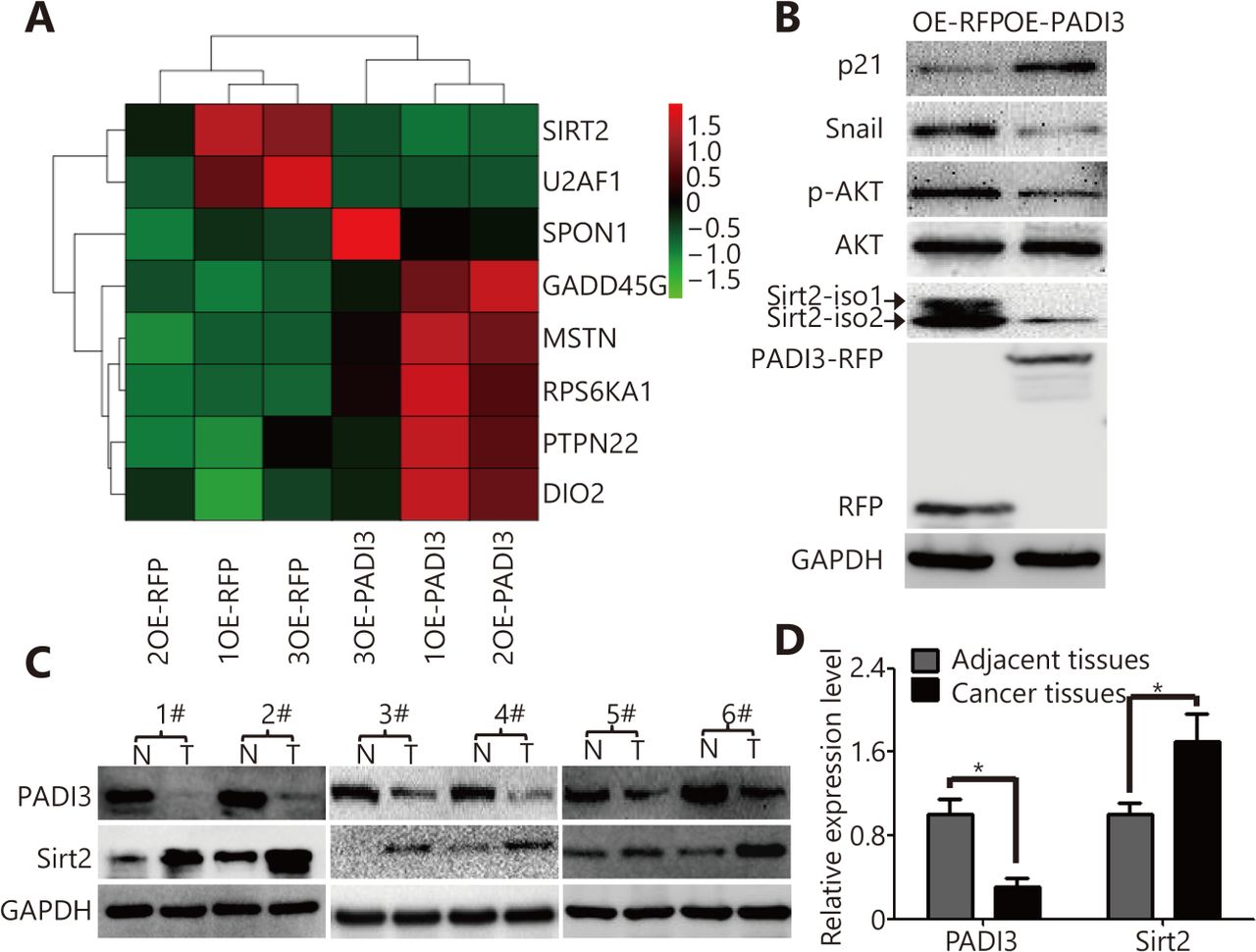
    RNA-sequencing analysis of PADI3-expressing HCT116 cells. (A) Heat map from the RNA-sequencing analysis. (B) Expression of phosphorylated AKT (p-AKT), AKT, Sirt2, Snail and p21 in OE-PADI3 HCT116 cells, OE-RFP HCT116 cells is selected as the control group. (C) Western blot analysis was used to measure the expression level of Sirt2 and PADI3 in colon cancer tissues and corresponding tissues. (D) Statistical analysis of the expression level of Sirt2 and PADI3. * indicates P < 0.05 for three independent experiments analyzed by Student’s t-test.

  • 6
    • Download figure
    • Open in new tab
    • Download powerpoint
    6

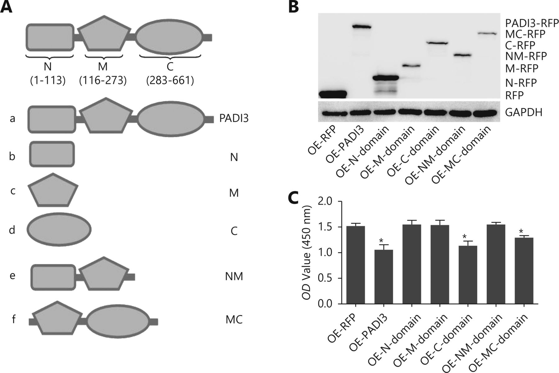
    Functional analysis of different PADI3 domains. (A) Predicting the PADI3-domain structure using SMART. (B) Western blot analysis was used to detect the expression of different PADI3 domains in HCT116 cells. (C) CCK-8 assay was used to measure the cell proliferation activity of different PADI3 domains. * indicates P < 0.05 for three independent experiments analyzed by Student’s t-test.

  • 7
    • Download figure
    • Open in new tab
    • Download powerpoint
    7

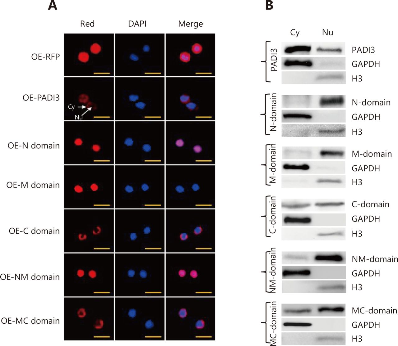
    Subcellular locations of the truncated forms of PADI3 in HCT116 cells. (A) Red fluorescence indicates RFP or PADI3-fused RFP, and blue indicates nuclei stained with DAPI. The merged image shows both RFP fluorescence and DAPI staining. The scale bar represents 20 µm. (B) Western blot analysis was used to detect truncated PADI3 in the cytoplasm and nucleus. GAPDH was used as the internal control for cytoplasmic expression, and histone 3 was used as the internal control for nuclear expression. Nu: nucleus, Cy: cytoplasm.

  • 8
    • Download figure
    • Open in new tab
    • Download powerpoint
    8

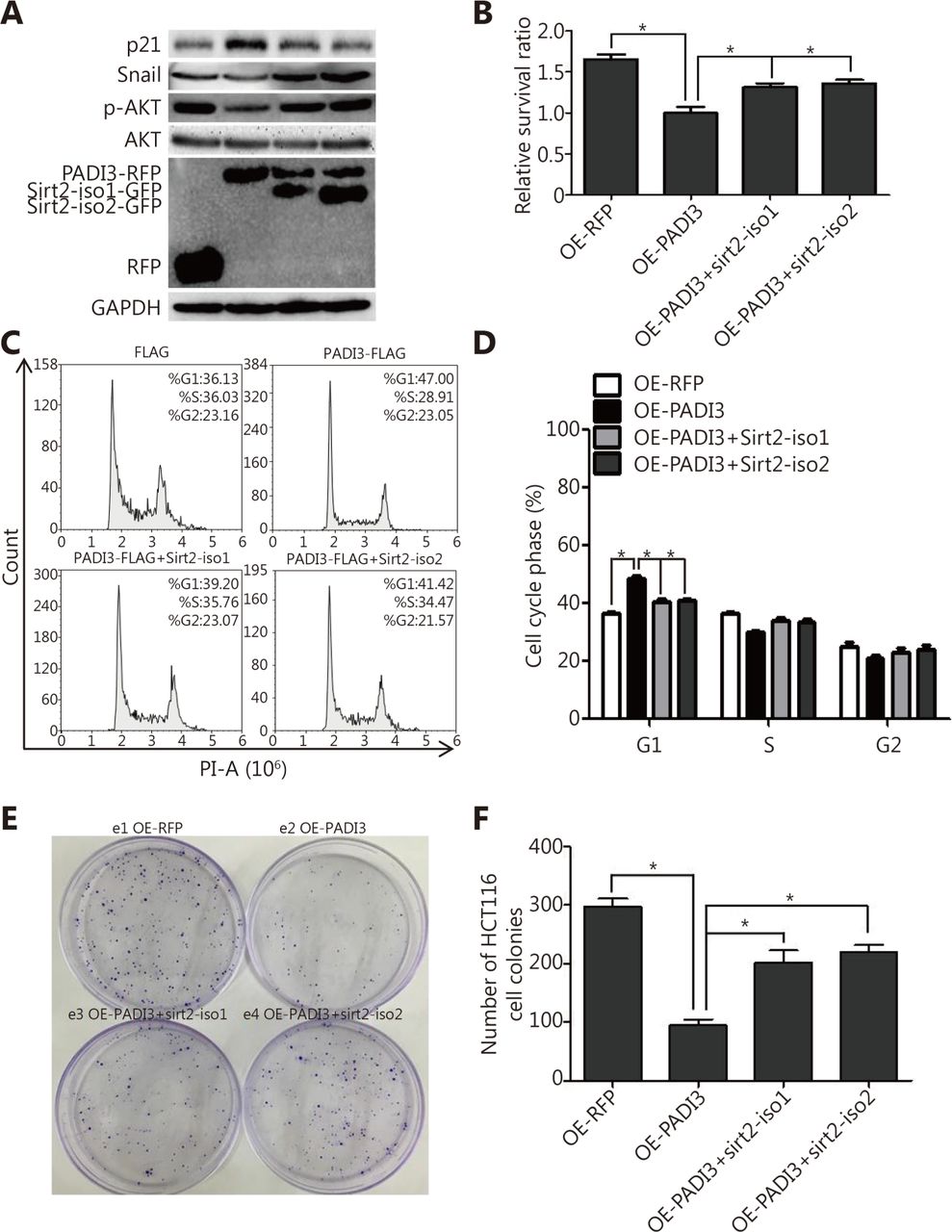
    Effects of Sirt2 on PADI3-expressing HCT116 cells. PADI3-expressing HCT116 cells were transfected with pCDNA3.1-Sirt2-iso1 plasmids (OE-PADI3-Sirt2-iso1) or pCDNA3.1-Sirt2-iso2 plasmids (OE-PADI3-Sirt2-iso2) separately. PADI3-expressing HCT116 cells were used as the positive control, and RFP-expressing HCT116 cells were used as the negative control. (A) Western blot analysis was used to measure the expression of AKT, p-AKT, Snail and p21. (B) A CCK-8 assay was used to measure cell proliferation. (C) The cell cycle was analyzed using FCM. (D) Statistical analysis of C. (E) Colony number and size were analyzed. (F) Statistical analysis of E. * indicates P < 0.05 for three independent experiments analyzed by Student’s t-test.

  • S1
    • Download figure
    • Open in new tab
    • Download powerpoint
    S1

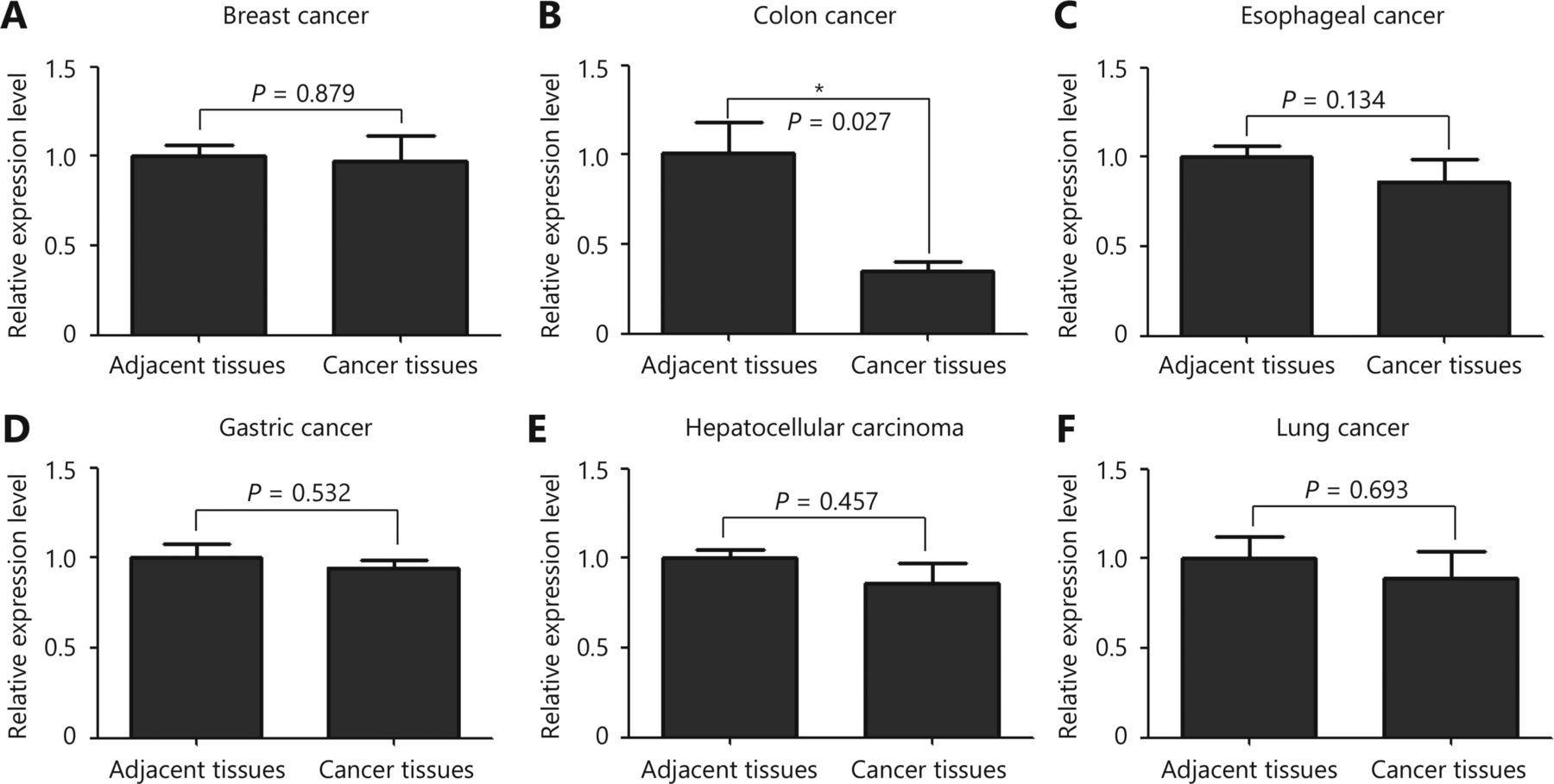
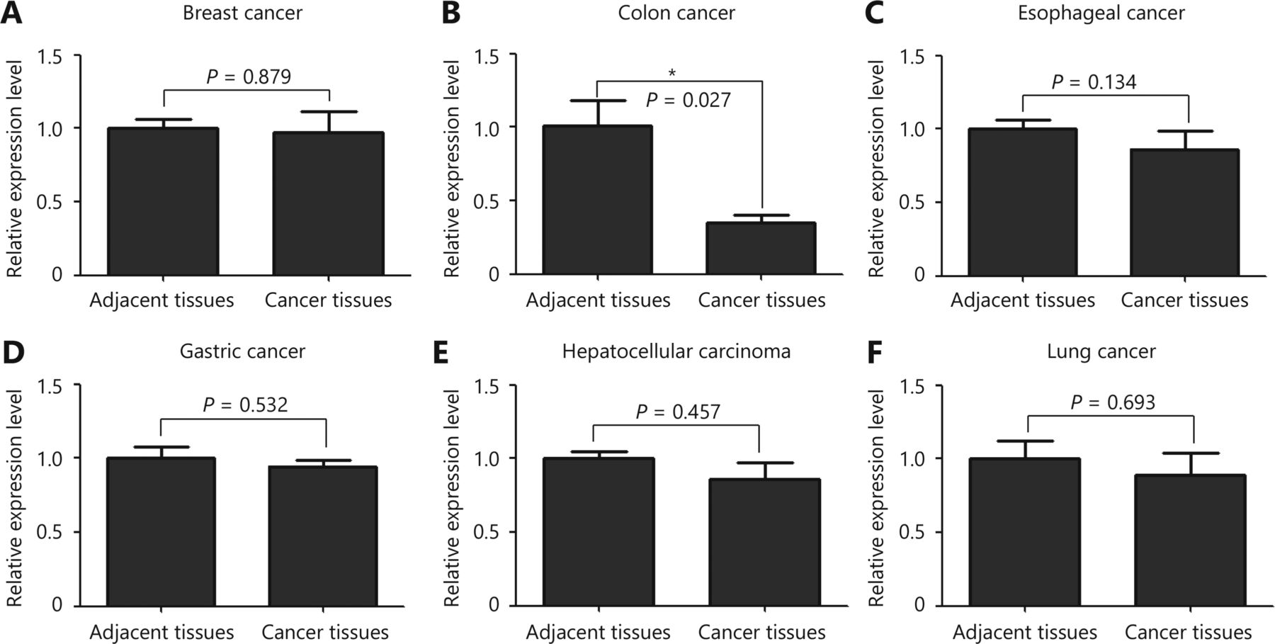
    PADI3 expression profile in various tumor tissues measured using qRT-PCR.
Tables

  • Figures
    • View popup
    S1

    PCR primer sequences

    PrimerSequence
    PADI3-QF:CCCTCGTGGACATTTATGGGT
    PADI3-QR:ATCTCCAAAGTCGCGTCAAAG
    GAPDH-QF:GCACCGTCAAGGCTGAGAAC
    GAPDH-QR:TGGTGAAGACGCCAGTGGA
    PADI3-EcoRI-Fex:TACTCAGAATTCATGTCGCTGCAGAGAATC
    PADI3-AscI-Rex:TACTCAGGCGCGCCGAGGGCACCATGTTCCACCA
    PADI3-N-OE-EcoRI-Fex:TACTCAGAATTCATGTCGCTGCAGAGAATC
    PADI3-N-OE-AscI-Rex:TACTCAGGCGCGCCGAGGTGAGGTAGAGCACCGC
    PADI3-M-OE-EcoRI-Fex:TACTCAGAATTCGTTGACATCTCTCTGGAT
    PADI3-M-OE-AscI-Rex:TACTCAGGCGCGCCGAGTCCAGCAGAGTGACATG
    PADI3-C-OE-EcoRI-Fex:TACTCAGAATTCCCTATCTTCACTGACACT
    PADI3-C-OE-AscI-Rex:TACTCAGGCGCGCCGAGTTCCACCACTTGAAAGA
    Sirt2-iso1-Fex:TACTCAGAATTCATGGCAGAGCCAGACCCC
    Sirt2-iso1-Rex:TACTCAGGCGCGCCGACTGGGGTTTCTCCCTCTC
    Sirt2-iso2-Fex:TACTCAGAATTCATGGACTTCCTGCGGAAC
    Sirt2-iso2-Rex:TACTCAGGCGCGCCGACTGGGGTTTCTCCCTCTC
PreviousNext
Back to top

In this issue

Cancer Biology and Medicine: 16 (4)
Cancer Biology & Medicine
Vol. 16, Issue 4
1 Nov 2019
  • Table of Contents
  • Index by author
Print
Download PDF
Email Article

Thank you for your interest in spreading the word on Cancer Biology & Medicine.

NOTE: We only request your email address so that the person you are recommending the page to knows that you wanted them to see it, and that it is not junk mail. We do not capture any email address.

Enter multiple addresses on separate lines or separate them with commas.
PADI3 induces cell cycle arrest via the Sirt2/AKT/p21 pathway and acts as a tumor suppressor gene in colon cancer
(Your Name) has sent you a message from Cancer Biology & Medicine
(Your Name) thought you would like to see the Cancer Biology & Medicine web site.
Citation Tools
PADI3 induces cell cycle arrest via the Sirt2/AKT/p21 pathway and acts as a tumor suppressor gene in colon cancer
Xiaotian Chang, Zhengbin Chai, Jiaorui Zou, Hongxing Wang, Yao Wang, Yabing Zheng, Hui Wu, Chunyan Liu
Cancer Biology & Medicine Nov 2019, 16 (4) 729-742; DOI: 10.20892/j.issn.2095-3941.2019.0065

Citation Manager Formats

  • BibTeX
  • Bookends
  • EasyBib
  • EndNote (tagged)
  • EndNote 8 (xml)
  • Medlars
  • Mendeley
  • Papers
  • RefWorks Tagged
  • Ref Manager
  • RIS
  • Zotero
Share
PADI3 induces cell cycle arrest via the Sirt2/AKT/p21 pathway and acts as a tumor suppressor gene in colon cancer
Xiaotian Chang, Zhengbin Chai, Jiaorui Zou, Hongxing Wang, Yao Wang, Yabing Zheng, Hui Wu, Chunyan Liu
Cancer Biology & Medicine Nov 2019, 16 (4) 729-742; DOI: 10.20892/j.issn.2095-3941.2019.0065
Twitter logo Facebook logo Mendeley logo
  • Tweet Widget
  • Facebook Like
  • Google Plus One

Jump to section

  • Article
    • Abstract
    • Introduction
    • Materials and methods
    • Results
    • Discussion
    • Conclusions
    • Acknowledgments
    • Conflict of interest statement
    • Supplementary materials
    • References
  • Figures & Data
  • Info & Metrics
  • References
  • PDF

Related Articles

  • No related articles found.
  • Google Scholar

Cited By...

  • Ca2+-mediated protein citrullination regulates proliferation in the regenerating and malignant CNS
  • Google Scholar

More in this TOC Section

  • Psychological health mediates the association between diet and upper gastrointestinal cancer: a cross-sectional analysis from a large-scale population-based screening project
  • Lung cancer mortality trends in China from 2013 to 2021 and projections to 2030
  • Global landscape and temporal trends in lifetime risk of colorectal cancer in 185 countries: a population-based study
Show more Original Article

Similar Articles

Keywords

  • PADI3
  • SIRT2
  • colon cancer
  • cell cycle
  • C-domain

Navigate

  • Home
  • Current Issue

More Information

  • About CBM
  • About CACA
  • About TMUCIH
  • Editorial Board
  • Subscription

For Authors

  • Instructions for authors
  • Journal Policies
  • Submit a Manuscript

Journal Services

  • Email Alerts
  • Facebook
  • RSS Feeds
  • Twitter


© 2026 Cancer Biology & Medicine

Powered by HighWire