
Main menu

  • Home
  • About
    • About CBM
    • Editorial Board
  • Articles
    • Ahead of print
    • Current Issue
    • Archive
    • Collections
    • Cover Story
  • For Authors
    • Instructions for Authors
    • Resources
    • Submit a Manuscript
  • For Reviewers
    • Become a Reviewer
    • Instructions for Reviewers
    • Resources
    • Outstanding Reviewer
  • Subscription
  • Alerts
    • Email Alerts
    • RSS Feeds
    • Table of Contents
  • Contact us
  • Other Publications
    • cbm

User menu

  • My alerts

Search

  • Advanced search
Cancer Biology & Medicine
  • Other Publications
    • cbm
  • My alerts
Cancer Biology & Medicine

Advanced Search


  • Home
  • About
    • About CBM
    • Editorial Board
  • Articles
    • Ahead of print
    • Current Issue
    • Archive
    • Collections
    • Cover Story
  • For Authors
    • Instructions for Authors
    • Resources
    • Submit a Manuscript
  • For Reviewers
    • Become a Reviewer
    • Instructions for Reviewers
    • Resources
    • Outstanding Reviewer
  • Subscription
  • Alerts
    • Email Alerts
    • RSS Feeds
    • Table of Contents
  • Contact us
  • Follow cbm on Twitter
  • Visit cbm on Facebook
OtherPerspective

Evolution-driven crosstalk between glioblastoma and the tumor microenvironment

Lingxiang Wu, Ruichao Chai, Zihan Lin, Rongrong Wu, Diru Yao, Tao Jiang and Qianghu Wang
Cancer Biology & Medicine March 2023, 20220771; DOI: https://doi.org/10.20892/j.issn.2095-3941.2022.0771
Lingxiang Wu
1Department of Bioinformatics, Nanjing Medical University, Nanjing 211166, China
2Institute for Brain Tumors, Jiangsu Collaborative Innovation Center for Cancer Personalized Medicine, Nanjing Medical University, Nanjing 211166, China
  • Find this author on Google Scholar
  • Find this author on PubMed
  • Search for this author on this site
Ruichao Chai
3Department of Molecular Neuropathology, Beijing Neurosurgical Institute, Capital Medical University, Beijing 100070, China
4Chinese Glioma Genome Atlas Network (CGGA), Beijing, China
5Department of Neurosurgery, Beijing Tiantan Hospital, Capital Medical University, Beijing 100070, China
  • Find this author on Google Scholar
  • Find this author on PubMed
  • Search for this author on this site
Zihan Lin
1Department of Bioinformatics, Nanjing Medical University, Nanjing 211166, China
2Institute for Brain Tumors, Jiangsu Collaborative Innovation Center for Cancer Personalized Medicine, Nanjing Medical University, Nanjing 211166, China
  • Find this author on Google Scholar
  • Find this author on PubMed
  • Search for this author on this site
Rongrong Wu
1Department of Bioinformatics, Nanjing Medical University, Nanjing 211166, China
2Institute for Brain Tumors, Jiangsu Collaborative Innovation Center for Cancer Personalized Medicine, Nanjing Medical University, Nanjing 211166, China
  • Find this author on Google Scholar
  • Find this author on PubMed
  • Search for this author on this site
Diru Yao
1Department of Bioinformatics, Nanjing Medical University, Nanjing 211166, China
2Institute for Brain Tumors, Jiangsu Collaborative Innovation Center for Cancer Personalized Medicine, Nanjing Medical University, Nanjing 211166, China
  • Find this author on Google Scholar
  • Find this author on PubMed
  • Search for this author on this site
Tao Jiang
3Department of Molecular Neuropathology, Beijing Neurosurgical Institute, Capital Medical University, Beijing 100070, China
4Chinese Glioma Genome Atlas Network (CGGA), Beijing, China
5Department of Neurosurgery, Beijing Tiantan Hospital, Capital Medical University, Beijing 100070, China
  • Find this author on Google Scholar
  • Find this author on PubMed
  • Search for this author on this site
  • ORCID record for Tao Jiang
  • For correspondence: [email protected] [email protected]
Qianghu Wang
1Department of Bioinformatics, Nanjing Medical University, Nanjing 211166, China
2Institute for Brain Tumors, Jiangsu Collaborative Innovation Center for Cancer Personalized Medicine, Nanjing Medical University, Nanjing 211166, China
  • Find this author on Google Scholar
  • Find this author on PubMed
  • Search for this author on this site
  • ORCID record for Qianghu Wang
  • For correspondence: [email protected] [email protected]
  • Article
  • Figures & Data
  • References
  • Info & Metrics
  • PDF
Loading

Glioblastoma (GBM) is a malignant adult brain tumor for which 90% of patients experience recurrence within a year after surgery1. Evolution confers treatment resistance capabilities on tumors2. The diversification of malignant and non-malignant (i.e., stromal and immune cell) compartments in the tumor microenvironment (TME) during tumor evolution3–7 eventually results in the formation of a complex interaction network that promotes tumor progression.

Tumor evolution endows GBM with the plasticity to adapt to environmental stresses

Numerous studies8–11 have demonstrated that GBM exhibits a variety of molecular states associated with specific genomic variation and TME characteristics. Using single-cell tracing technology, Neftel et al.11 have demonstrated that tumor cells in a single state can form a tumor block consisting of multiple cellular states after being implanted in mice, thus indicating that these tumor cells have high plasticity and can dynamically transform cell states. Our recent study12 has indicated that the traits formed during natural tumor evolution are associated with the dynamic maintenance of these tumor states. During long-term evolution, tumor cells simultaneously activate several transcription factors associated with stemness and differentiation phenotypes, including SERPINH1 and ATF513–15. Tumor cells with a high natural evolution signature, after injection into the brains of mice, have been found to regenerate a variety of cell states in the tissues, thereby increasing heterogeneity.

The high plasticity conferred by tumor evolution indicates that tumor cells can adapt to TME changes. Poor blood flow in the central necrotic areas of tumors creates a hypoxic environment. Our study12 has demonstrated that tumor cells activate the HIF1A/FOSL2 (AP-1 transcription factor subunit) regulatory axis under hypoxic conditions, thereby switching tumor states and remodeling the immunosuppressive microenvironment to support tumor development. GBM has also been found to induce WNT5A to mediate tumor cell differentiation into endothelium-like cells via the PAX6/DLX5 transcription program, and subsequently recruit peripheral endothelial cells, which form pseudo-blood vessels, thereby facilitating tumor-invasive growth16.

In response to treatment pressure, such as radiotherapy, tumor cells in the subventricular area have been found to activate an internal signaling pathway by binding CXCL12 in the microenvironment, thereby promoting the transformation of tumor cells into a mesenchymal-like state and facilitating their survival17. Such a state transition has recently been further confirmed by Wang et al.18, whose analysis of 86 primary-recurrent patient-paired GBM specimens with single-cell sequencing data has demonstrated that relapsed GBM is characterized by a shift to a mesenchymal-like state, which is mediated by AP-1. In addition, we have recently found that PTPRZ1-MET fusion and a high-frequency of MET exon 14 skipping (METex14) occur in brain metastases of lung cancer19. We also found that METex14 is present in a higher proportion (14%) of secondary glioblastomas than pan-gliomas (0.4%)2, thus suggesting that tumors bearing METex14 tend to occur in the brain microenvironment in advanced tumors. Consequently, these findings imply co-evolution of the tumor and microenvironment.

Tumor evolution reconstructs the TME interaction network

The interaction between tumors and the TME evolves in tandem with tumor evolution (Figure 1). In a healthy brain, glial cells (oligodendrocytes, microglia, and astrocytes) perform various functions, such as axon myelination, immune surveillance, and blood-brain barrier (BBB) maintenance20. In the early stages of GBM evolution, microglia make up most macrophages and have proinflammatory roles through expressing cytokines such as TNF-α and IL-1β21, thus implying that they might play a role in activating inflammation and fighting tumor cells. However, evidence suggests that the IL-1β released by microglia binds the IL-1 receptor (IL-1R) on the surfaces of tumor cells and activates downstream pathways that promote tumor cell proliferation22. Yeo et al.21 have speculated that GBM stimulates the production and differentiation of brain resident oligodendrocytes. These oligodendrocytes in turn promote the invasiveness of GBM cells via the angiopoietin-2 signaling pathway23. Astrocytes also facilitate tumor cell infiltration by activating zinc finger E-box-binding homeobox 1 (ZEB1)24. Furthermore, astrocytes secrete proteases (uPA) that increase the expression of matrix metalloproteinases released by GBM cells, thus leading to extracellular matrix remodeling and promoting invasion25. Moreover, Venkataramani et al.26 have revealed that neurons have a typical synaptic ultrastructure located on tumor microtubes that generate postsynaptic currents mediated by AMPA subtype glutamate receptors on tumor cells, thereby promoting tumor growth and invasion.

Figure 1
  • Download figure
  • Open in new tab
  • Download powerpoint
Figure 1

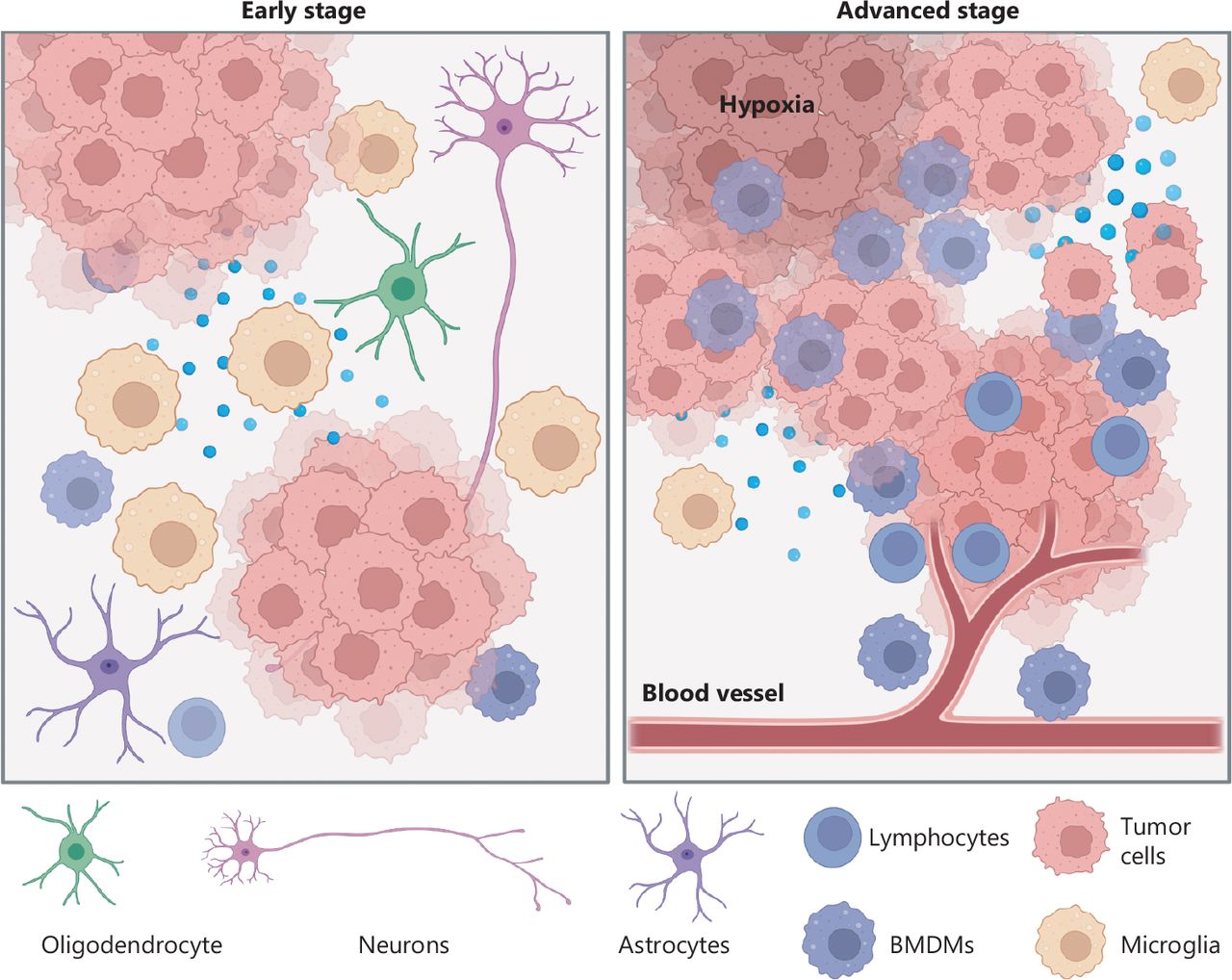
Coevolution of tumors and the tumor microenvironment. In early stages, tumor cells are often surrounded by neurons and glial cells. Crosstalk between tumors and the tumor microenvironment endows tumor cells with proliferation and invasion capabilities. In advanced stages, increased numbers of peripheral cells (e.g., BMDMs, lymphocytes, etc.) invade the tumor tissue. The high infiltration of BMDMs produces an immunosuppressive environment that further promotes tumor progression. Furthermore, hypoxic stress also accelerates tumor evolution.

The BBB, formed by the interplay of astrocyte foot processes with endothelial cells and pericytes, tightly regulates the inflow and outflow of substances to maintain a homeostatic environment for proper brain functioning27,28. However, tumor evolution may impair the BBB via inflammation, physical distortion, or increased vascularity, thus contributing to blood vessel leakage29. Consequently, many peripheral blood cells invade tumor tissue. Recent studies12,21 have confirmed that high infiltration of bone-marrow-derived macrophages (BMDMs) is associated with advanced evolution of GBM. Tumor cells may educate these BMDMs with multiple factors (e.g., CSF1), thus eventually forming an immunosuppressive microenvironment that inhibits T cell proliferation and interferon-gamma production. Other immune cells, such as regulatory T cells (Tregs), are also observed in high numbers in advanced stages of GBM progression30. Tregs are recruited to tumor tissue by CCL2/CCL22 and subsequently promote GBM progression. Tregs also inhibit the activity of T cells, and exert antitumor effects by secreting the cytokines IL-10 and TGF-β31. Furthermore, Zhang et al.32 have demonstrated that pericytes derived from the peripheral blood interact with tumor cells regardless of vascular structure. These pericytes activate the DNA damage repair pathway by secreting CCL5, which binds CCR5 on tumor cells, thus rendering glioma cells resistant to temozolomide.

Current GBM evolutionary research strategies

Numerous studies have been conducted to deconvolve tumor evolution by investigating the characteristics of GBM (Table 1). Primary and recurrent GBM longitudinal cohorts are the classical model widely used to examine for tumor evolution driven by treatment4,6,18,33–35. For instance, Wang et al.4 have assessed the longitudinal genome and transcriptomic data of 114 patients, and have found that GBM has a highly branched evolutionary pattern during its recurrence: 63% of the patients experienced changes in GBM molecular subtypes. These studies have substantially deepened understanding of the tumor progression patterns driven by various treatments and have provided essential guidelines for the clinical management of recurrent GBM.

View this table:
  • View inline
  • View popup
Table 1

Strategies for exploring tumor evolution

Ozawa et al.36 have predicted the variation order in GBM through data modeling of massive GBM genomic variation, thus uncovering features in the natural evolution of primary GBM. Compared with other genomic alterations, chromosome 7 amplification and chromosome 10 deletion occur early in tumor evolution. Korber et al.37 have used a similar strategy to confirm this finding. Furthermore, their analysis has revealed that mutation of the TERT promoter is a prerequisite for rapid tumor growth. Although these computational methods provide powerful tools for dissecting driver events in tumor evolution, focusing on tumor genomic traits may fail to interrogate the dynamic changes in the microenvironment.

Multiple lesions from the same clonal ancestor can develop in a time- and space-independent manner, thus providing snapshots that reflect the different stages of tumor evolution12,38–40. Abou-El-Ardat et al.38 have found that, in advanced stages of GBM, aberrations in the RTK/PI3K, p53, and RB regulatory pathways are common and may be the factors driving subcloning. Our recent study has further examined multifocal GBMs with single-cell RNA sequencing and identified a positive feedback interaction between tumor cells and macrophages during GBM evolution12. Tumor cells in advanced stages of evolution recruit and polarize macrophages through ANXA1. These recruited BMDMs then secrete cytokines that promote tumor invasion and evolution.

Other strategies have leveraged the advantages of organoids41–43 or animal models21,44,45. Tang et al.41 have developed a rapid three-dimensional bioprinting method to integrate tumor cells and other stroma and immune cells for co-culture; this method may serve as a platform for studying cellular crosstalk during tumor evolution. Yeo et al.21 have used a mouse GBM model and single-cell RNA sequencing to characterize the multifaceted roles of macrophages from early to late stages of GBM evolution.

Future perspectives

Owing to the inherent differences in the microenvironment among tumor regions, the heterogeneity of tumor cells among these regions is particularly pronounced. Puchalski et al.46 have constructed an anatomical transcription map of GBM and have found that mesenchymal-like tumor cells are enriched mainly in hypoxic and necrotic areas. In contrast, classical-like tumor cells are associated with vascular proliferation and infiltration areas. Thus, this preference for tumor cell enrichment could eventually result in a distinct crosstalk pattern between tumors and the tumor microenvironment across tumor regions. Future research should incorporate spatial factors into the interactions to thoroughly investigate cell communication, particularly that mediated by paracrine and direct signaling.

Beyond cell-to-cell communication, metabolism has been found to be an important factor in regulating interaction networks47. Altered metabolism in the tumor microenvironment can lead to the production of extracellular metabolites that alter the states and phenotypes of various cells. For instance, the GBM-derived metabolite KYN is involved in switching TAMs from the M1 to the M2 phenotype by activating the transcription factor AHR48. Glycolysis has been found to alter the microenvironment by producing lactic acid, which regulates T cell immune activity49. Consequently, dissecting the components of metabolites in the microenvironment will be crucial for establishing the potential links between tumors and the tumor microenvironment.

Given the spatial heterogeneity and dynamic changes in the tumor microenvironment alongside tumor evolution, the interplay between tumors and the tumor microenvironment is complex and regulated by multiple factors, including stresses, etc. Advances in single-cell technologies, such as single-cell tracking technology50 and single-cell spatial omics51,52, have led to potential clues for in-depth analysis of tumor-TME coevolution, and may contribute to better understanding of the mechanisms underlying cancer evolution.

Conflict of interest statement

No potential conflicts of interest are disclosed.

Footnotes

  • ↵*These authors contributed equally to this work.

  • Received January 1, 2023.
  • Accepted January 10, 2023.
  • Copyright: © 2023, Cancer Biology & Medicine
https://creativecommons.org/licenses/by/4.0/

This is an open access article distributed under the terms of the Creative Commons Attribution License (CC BY) 4.0, which permits unrestricted use, distribution and reproduction in any medium, provided the original author and source are credited.

References

  1. 1.↵
    1. Jiang T,
    2. Nam DH,
    3. Ram Z,
    4. Poon WS,
    5. Wang J,
    6. Boldbaatar D, et al.
    Clinical practice guidelines for the management of adult diffuse gliomas. Cancer Lett. 2021; 499: 60–72.
    OpenUrl
  2. 2.↵
    1. Hu H,
    2. Mu Q,
    3. Bao Z,
    4. Chen Y,
    5. Liu Y,
    6. Chen J, et al.
    Mutational landscape of secondary glioblastoma guides MET-targeted trial in brain tumor. Cell. 2018; 175: 1665–78.e18.
    OpenUrlCrossRefPubMed
  3. 3.↵
    1. Kim J,
    2. Lee IH,
    3. Cho HJ,
    4. Park CK,
    5. Jung YS,
    6. Kim Y, et al.
    Spatiotemporal evolution of the primary glioblastoma genome. Cancer Cell. 2015; 28: 318–28.
    OpenUrlCrossRefPubMed
  4. 4.↵
    1. Wang J,
    2. Cazzato E,
    3. Ladewig E,
    4. Frattini V,
    5. Rosenbloom DI,
    6. Zairis S, et al.
    Clonal evolution of glioblastoma under therapy. Nat Genet. 2016; 48: 768–76.
    OpenUrlCrossRefPubMed
  5. 5.
    1. Barthel FP,
    2. Johnson KC,
    3. Varn FS,
    4. Moskalik AD,
    5. Tanner G,
    6. Kocakavuk E, et al.
    Longitudinal molecular trajectories of diffuse glioma in adults. Nature. 2019; 576: 112–20.
    OpenUrlCrossRefPubMed
  6. 6.↵
    1. Kim H,
    2. Zheng S,
    3. Amini SS,
    4. Virk SM,
    5. Mikkelsen T,
    6. Brat DJ, et al.
    Whole-genome and multisector exome sequencing of primary and post-treatment glioblastoma reveals patterns of tumor evolution. Genome Res. 2015; 25: 316–27.
    OpenUrlAbstract/FREE Full Text
  7. 7.↵
    1. Wu L,
    2. Chai R,
    3. Zhao Z,
    4. Wang Q,
    5. Jiang T.
    Role of the tumor microenvironment in shaping IDH-wildtype glioma plasticity, and potential therapeutic strategies. Cancer Biol Med. 2022; 19: 1423–7.
    OpenUrlFREE Full Text
  8. 8.↵
    1. Verhaak RG,
    2. Hoadley KA,
    3. Purdom E,
    4. Wang V,
    5. Qi Y,
    6. Wilkerson MD, et al.
    Integrated genomic analysis identifies clinically relevant subtypes of glioblastoma characterized by abnormalities in PDGFRA, IDH1, EGFR, and NF1. Cancer Cell. 2010; 17: 98–110.
    OpenUrlCrossRefPubMed
  9. 9.
    1. Wang Q,
    2. Hu B,
    3. Hu X,
    4. Kim H,
    5. Squatrito M,
    6. Scarpace L, et al.
    Tumor evolution of glioma-intrinsic gene expression subtypes associates with immunological changes in the microenvironment. Cancer Cell. 2017; 32: 42–56.e6.
    OpenUrlCrossRefPubMed
  10. 10.
    1. Garofano L,
    2. Migliozzi S,
    3. Oh YT,
    4. D’Angelo F,
    5. Najac RD,
    6. Ko A, et al.
    Pathway-based classification of glioblastoma uncovers a mitochondrial subtype with therapeutic vulnerabilities. Nat Cancer. 2021; 2: 141–56.
    OpenUrl
  11. 11.↵
    1. Neftel C,
    2. Laffy J,
    3. Filbin MG,
    4. Hara T,
    5. Shore ME,
    6. Rahme GJ, et al.
    An integrative model of cellular states, plasticity, and genetics for glioblastoma. Cell. 2019; 178: 835–49.
    OpenUrlPubMed
  12. 12.↵
    1. Wu L,
    2. Wu W,
    3. Zhang J,
    4. Zhao Z,
    5. Li L,
    6. Zhu M, et al.
    Natural coevolution of tumor and immunoenvironment in glioblastoma. Cancer Discov. 2022; 12: 2820–37.
    OpenUrl
  13. 13.↵
    1. Xiong G,
    2. Chen J,
    3. Zhang G,
    4. Wang S,
    5. Kawasaki K,
    6. Zhu J, et al.
    Hsp47 promotes cancer metastasis by enhancing collagen-dependent cancer cell-platelet interaction. Proc Natl Acad Sci U S A. 2020; 117: 3748–58.
    OpenUrlAbstract/FREE Full Text
  14. 14.
    1. Jiang X,
    2. Zhou T,
    3. Wang Z,
    4. Qi B,
    5. Xia H.
    HSP47 Promotes glioblastoma stemlike cell survival by modulating tumor microenvironment extracellular matrix through TGF-β pathway. ACS Chem Neurosci. 2017; 8: 128–34.
    OpenUrlCrossRefPubMed
  15. 15.↵
    1. Wang SZ,
    2. Ou J,
    3. Zhu LJ,
    4. Green MR.
    Transcription factor ATF5 is required for terminal differentiation and survival of olfactory sensory neurons. Proc Natl Acad Sci U S A. 2012; 109: 18589–94.
    OpenUrlAbstract/FREE Full Text
  16. 16.↵
    1. Hu B,
    2. Wang Q,
    3. Wang YA,
    4. Hua S,
    5. Sauvé CG,
    6. Ong D, et al.
    Epigenetic activation of WNT5A drives glioblastoma stem cell differentiation and invasive growth. Cell. 2016; 167: 1281–95.e18.
    OpenUrlCrossRef
  17. 17.↵
    1. Goffart N,
    2. Lombard A,
    3. Lallemand F,
    4. Kroonen J,
    5. Nassen J,
    6. Di Valentin E, et al.
    CXCL12 mediates glioblastoma resistance to radiotherapy in the subventricular zone. Neuro Oncol. 2017; 19: 66–77.
    OpenUrlCrossRefPubMed
  18. 18.↵
    1. Wang L,
    2. Jung J,
    3. Babikir H,
    4. Shamardani K,
    5. Jain S,
    6. Feng X, et al.
    A single-cell atlas of glioblastoma evolution under therapy reveals cell-intrinsic and cell-extrinsic therapeutic targets. Nat Cancer. 2022; 3: 1534–52.
    OpenUrl
  19. 19.↵
    1. Chai RC,
    2. Liu X,
    3. Pang B,
    4. Liu YQ,
    5. Li JJ,
    6. Li YF, et al.
    Recurrent PTPRZ1-MET fusion and a high occurrence rate of MET exon 14 skipping in brain metastases. Cancer Sci. 2022; 113: 796–801.
    OpenUrl
  20. 20.↵
    1. Jakel S,
    2. Dimou L.
    Glial cells and their function in the adult brain: a journey through the history of their ablation. Front Cell Neurosci. 2017; 11: 24.
    OpenUrlCrossRef
  21. 21.↵
    1. Yeo AT,
    2. Rawal S,
    3. Delcuze B,
    4. Christofides A,
    5. Atayde A,
    6. Strauss L, et al.
    Single-cell RNA sequencing reveals evolution of immune landscape during glioblastoma progression. Nat Immunol. 2022; 23: 971–84.
    OpenUrl
  22. 22.↵
    1. Liu H,
    2. Sun Y,
    3. Zhang Q,
    4. Jin W,
    5. Gordon RE,
    6. Zhang Y, et al.
    Pro-inflammatory and proliferative microglia drive progression of glioblastoma. Cell Rep. 2021; 36: 109718.
    OpenUrlCrossRef
  23. 23.↵
    1. Kawashima T,
    2. Yashiro M,
    3. Kasashima H,
    4. Terakawa Y,
    5. Uda T,
    6. Nakajo K, et al.
    Oligodendrocytes up-regulate the invasive activity of glioblastoma cells via the angiopoietin-2 signaling pathway. Anticancer Res. 2019; 39: 577–84.
    OpenUrlAbstract/FREE Full Text
  24. 24.↵
    1. Iser IC,
    2. Lenz G,
    3. Wink MR.
    EMT-like process in glioblastomas and reactive astrocytes. Neurochem Int. 2019; 122: 139–43.
    OpenUrlCrossRef
  25. 25.↵
    1. Brandao M,
    2. Simon T,
    3. Critchley G,
    4. Giamas G.
    Astrocytes, the rising stars of the glioblastoma microenvironment. Glia. 2019; 67: 779–90.
    OpenUrl
  26. 26.↵
    1. Venkataramani V,
    2. Tanev DI,
    3. Strahle C,
    4. Studier-Fischer A,
    5. Fankhauser L,
    6. Kessler T, et al.
    Glutamatergic synaptic input to glioma cells drives brain tumour progression. Nature. 2019; 573: 532–8.
    OpenUrlCrossRefPubMed
  27. 27.↵
    1. Daneman R,
    2. Prat A.
    The blood-brain barrier. Cold Spring Harb Perspect Biol. 2015; 7: a020412.
  28. 28.↵
    1. Langen UH,
    2. Ayloo S,
    3. Gu C.
    Development and cell biology of the blood-brain barrier. Annu Rev Cell Dev Biol. 2019; 35: 591–613.
    OpenUrlCrossRef
  29. 29.↵
    1. Dubois LG,
    2. Campanati L,
    3. Righy C,
    4. D’Andrea-Meira I,
    5. Spohr TC,
    6. Porto-Carreiro I, et al.
    Gliomas and the vascular fragility of the blood brain barrier. Front Cell Neurosci. 2014; 8: 418.
    OpenUrlPubMed
  30. 30.↵
    1. Amoozgar Z,
    2. Kloepper J,
    3. Ren J,
    4. Tay RE,
    5. Kazer SW,
    6. Kiner E, et al.
    Targeting Treg cells with GITR activation alleviates resistance to immunotherapy in murine glioblastomas. Nat Commun. 2021; 12: 2582.
    OpenUrlCrossRef
  31. 31.↵
    1. Wang H,
    2. Zhou H,
    3. Xu J,
    4. Lu Y,
    5. Ji X,
    6. Yao Y, et al.
    Different T-cell subsets in glioblastoma multiforme and targeted immunotherapy. Cancer Lett. 2021; 496: 134–43.
    OpenUrl
  32. 32.↵
    1. Zhang XN,
    2. Yang KD,
    3. Chen C,
    4. He ZC,
    5. Wang QH,
    6. Feng H, et al.
    Pericytes augment glioblastoma cell resistance to temozolomide through CCL5-CCR5 paracrine signaling. Cell Res. 2021; 31: 1072–87.
    OpenUrl
  33. 33.↵
    1. Johnson BE,
    2. Mazor T,
    3. Hong C,
    4. Barnes M,
    5. Aihara K,
    6. McLean CY, et al.
    Mutational analysis reveals the origin and therapy-driven evolution of recurrent glioma. Science. 2014; 343: 189–93.
    OpenUrlAbstract/FREE Full Text
  34. 34.
    1. Osuka S,
    2. Van Meir EG.
    Overcoming therapeutic resistance in glioblastoma: the way forward. J Clin Invest. 2017; 127: 415–26.
    OpenUrlCrossRefPubMed
  35. 35.↵
    1. Varn FS,
    2. Johnson KC,
    3. Martinek J,
    4. Huse JT,
    5. Nasrallah MP,
    6. Wesseling P, et al.
    Glioma progression is shaped by genetic evolution and microenvironment interactions. Cell. 2022; 185: 2184–99.e16.
    OpenUrlCrossRef
  36. 36.↵
    1. Ozawa T,
    2. Riester M,
    3. Cheng YK,
    4. Huse JT,
    5. Squatrito M,
    6. Helmy K, et al.
    Most human non-GCIMP glioblastoma subtypes evolve from a common proneural-like precursor glioma. Cancer Cell. 2014; 26: 288–300.
    OpenUrlCrossRefPubMed
  37. 37.↵
    1. Korber V,
    2. Yang J,
    3. Barah P,
    4. Wu Y,
    5. Stichel D,
    6. Gu Z, et al.
    Evolutionary trajectories of IDH(WT) glioblastomas reveal a common path of early tumorigenesis instigated years ahead of initial diagnosis. Cancer Cell. 2019; 35: 692–704.e12.
    OpenUrl
  38. 38.↵
    1. Abou-El-Ardat K,
    2. Seifert M,
    3. Becker K,
    4. Eisenreich S,
    5. Lehmann M,
    6. Hackmann K, et al.
    Comprehensive molecular characterization of multifocal glioblastoma proves its monoclonal origin and reveals novel insights into clonal evolution and heterogeneity of glioblastomas. Neuro Oncol. 2017; 19: 546–57.
    OpenUrlCrossRef
  39. 39.
    1. Liu Q,
    2. Liu Y,
    3. Li W,
    4. Wang X,
    5. Sawaya R,
    6. Lang FF, et al.
    Genetic, epigenetic, and molecular landscapes of multifocal and multicentric glioblastoma. Acta Neuropathol. 2015; 130: 587–97.
    OpenUrlCrossRefPubMed
  40. 40.↵
    1. Lee JK,
    2. Wang J,
    3. Sa JK,
    4. Ladewig E,
    5. Lee HO,
    6. Lee IH, et al.
    Spatiotemporal genomic architecture informs precision oncology in glioblastoma. Nat Genet. 2017; 49: 594–9.
    OpenUrlCrossRefPubMed
  41. 41.↵
    1. Tang M,
    2. Xie Q,
    3. Gimple RC,
    4. Zhong Z,
    5. Tam T,
    6. Tian J, et al.
    Three-dimensional bioprinted glioblastoma microenvironments model cellular dependencies and immune interactions. Cell Res. 2020; 30: 833–53.
    OpenUrl
  42. 42.
    1. Zhang C,
    2. Jin M,
    3. Zhao J,
    4. Chen J,
    5. Jin W.
    Organoid models of glioblastoma: advances, applications and challenges. Am J Cancer Res. 2020; 10: 2242–57.
    OpenUrl
  43. 43.↵
    1. Jacob F,
    2. Salinas RD,
    3. Zhang DY,
    4. Nguyen PTT,
    5. Schnoll JG,
    6. Wong SZH, et al.
    A patient-derived glioblastoma organoid model and biobank recapitulates inter- and intra-tumoral heterogeneity. Cell. 2020; 180: 188–204.
    OpenUrlCrossRefPubMed
  44. 44.↵
    1. Tentler JJ,
    2. Tan AC,
    3. Weekes CD,
    4. Jimeno A,
    5. Leong S,
    6. Pitts TM, et al.
    Patient-derived tumour xenografts as models for oncology drug development. Nat Rev Clin Oncol. 2012; 9: 338–50.
    OpenUrlCrossRefPubMed
  45. 45.↵
    1. Miyai M,
    2. Tomita H,
    3. Soeda A,
    4. Yano H,
    5. Iwama T,
    6. Hara A.
    Current trends in mouse models of glioblastoma. J Neurooncol. 2017; 135: 423–32.
    OpenUrlCrossRefPubMed
  46. 46.↵
    1. Puchalski RB,
    2. Shah N,
    3. Miller J,
    4. Dalley R,
    5. Nomura SR,
    6. Yoon JG, et al.
    An anatomic transcriptional atlas of human glioblastoma. Science. 2018; 360: 660–3.
    OpenUrlAbstract/FREE Full Text
  47. 47.↵
    1. Carmona-Fontaine C,
    2. Deforet M,
    3. Akkari L,
    4. Thompson CB,
    5. Joyce JA,
    6. Xavier JB.
    Metabolic origins of spatial organization in the tumor microenvironment. Proc Natl Acad Sci U S A. 2017; 114: 2934–9.
    OpenUrlAbstract/FREE Full Text
  48. 48.↵
    1. Takenaka MC,
    2. Gabriely G,
    3. Rothhammer V,
    4. Mascanfroni ID,
    5. Wheeler MA,
    6. Chao CC, et al.
    Control of tumor-associated macrophages and T cells in glioblastoma via AHR and CD39. Nat Neurosci. 2019; 22: 729–40.
    OpenUrlCrossRefPubMed
  49. 49.↵
    1. Cascone T,
    2. McKenzie JA,
    3. Mbofung RM,
    4. Punt S,
    5. Wang Z,
    6. Xu C, et al.
    Increased tumor glycolysis characterizes immune resistance to adoptive T cell therapy. Cell Metab. 2018; 27: 977–87.e4.
    OpenUrlCrossRefPubMed
  50. 50.↵
    1. Wagner DE,
    2. Klein AM.
    Lineage tracing meets single-cell omics: opportunities and challenges. Nat Rev Genet. 2020; 21: 410–27.
    OpenUrl
  51. 51.↵
    1. Longo SK,
    2. Guo MG,
    3. Ji AL,
    4. Khavari PA.
    Integrating single-cell and spatial transcriptomics to elucidate intercellular tissue dynamics. Nat Rev Genet. 2021; 22: 627–44.
    OpenUrlCrossRefPubMed
  52. 52.↵
    1. Ma A,
    2. McDermaid A,
    3. Xu J,
    4. Chang Y,
    5. Ma Q.
    Integrative methods and practical challenges for single-cell multi-omics. Trends Biotechnol. 2020; 38: 1007–22.
    OpenUrl
Previous
Back to top

In this issue

Cancer Biology & Medicine: 20 (2)
Cancer Biology & Medicine
Vol. 20, Issue 2
15 Feb 2023
  • Table of Contents
  • Index by author
Print
Download PDF
Email Article

Thank you for your interest in spreading the word on Cancer Biology & Medicine.

NOTE: We only request your email address so that the person you are recommending the page to knows that you wanted them to see it, and that it is not junk mail. We do not capture any email address.

Enter multiple addresses on separate lines or separate them with commas.
Evolution-driven crosstalk between glioblastoma and the tumor microenvironment
(Your Name) has sent you a message from Cancer Biology & Medicine
(Your Name) thought you would like to see the Cancer Biology & Medicine web site.
Citation Tools
Evolution-driven crosstalk between glioblastoma and the tumor microenvironment
Lingxiang Wu, Ruichao Chai, Zihan Lin, Rongrong Wu, Diru Yao, Tao Jiang, Qianghu Wang
Cancer Biology & Medicine Mar 2023, 20220771; DOI: 10.20892/j.issn.2095-3941.2022.0771

Citation Manager Formats

  • BibTeX
  • Bookends
  • EasyBib
  • EndNote (tagged)
  • EndNote 8 (xml)
  • Medlars
  • Mendeley
  • Papers
  • RefWorks Tagged
  • Ref Manager
  • RIS
  • Zotero
Share
Evolution-driven crosstalk between glioblastoma and the tumor microenvironment
Lingxiang Wu, Ruichao Chai, Zihan Lin, Rongrong Wu, Diru Yao, Tao Jiang, Qianghu Wang
Cancer Biology & Medicine Mar 2023, 20220771; DOI: 10.20892/j.issn.2095-3941.2022.0771
Reddit logo Twitter logo Facebook logo Mendeley logo
  • Tweet Widget
  • Facebook Like
  • Google Plus One

Jump to section

  • Article
    • Tumor evolution endows GBM with the plasticity to adapt to environmental stresses
    • Tumor evolution reconstructs the TME interaction network
    • Current GBM evolutionary research strategies
    • Future perspectives
    • Conflict of interest statement
    • Footnotes
    • References
  • Figures & Data
  • Info & Metrics
  • References
  • PDF

Related Articles

  • No related articles found.
  • Google Scholar

Cited By...

  • No citing articles found.
  • Google Scholar

More in this TOC Section

  • Conceptualizing the complexity of ferroptosis to treat triple-negative breast cancer: theory-to-practice
  • Intercellular transmission of cGAS-STING signaling in cancer
Show more Perspective

Similar Articles

Navigate

  • Home
  • Current Issue

More Information

  • About CBM
  • About CACA
  • About TMUCIH
  • Editorial Board
  • Subscription

For Authors

  • Instructions for authors
  • Journal Policies
  • Submit a Manuscript

Journal Services

  • Email Alerts
  • Facebook
  • RSS Feeds
  • Twitter


© 2023 Cancer Biology & Medicine

Powered by HighWire