

Article Figures & Data
Figures
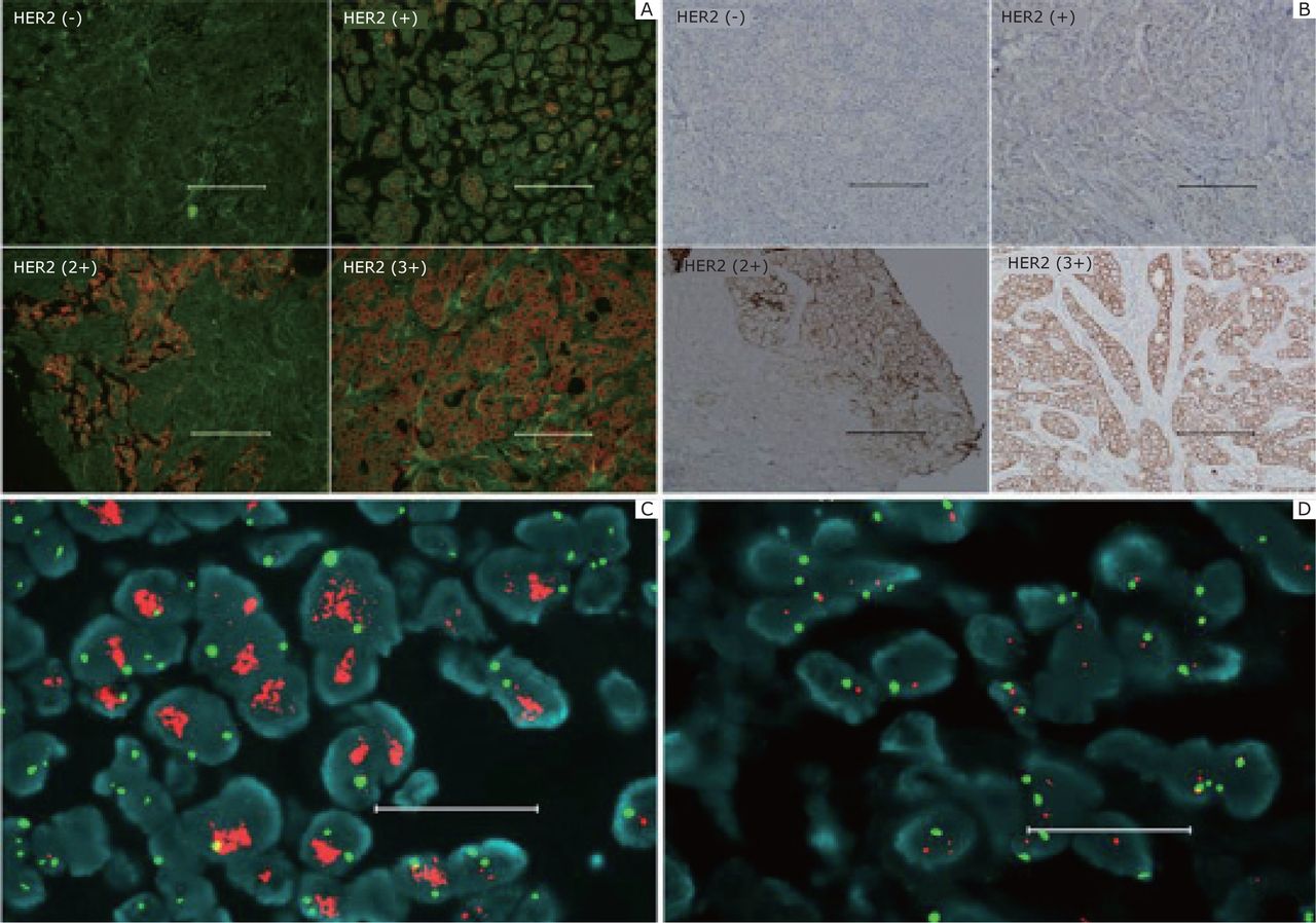
- 1
Accurate HER2 testing by QD–IHC. A: Specimens with different HER2 IHC scores detected by QD–IHC. B: Control for (A) by conventional IHC. C: FISH positive. D: Negative. Scale bar: 100 µm for (A) and (B); 20 µm for (C) and (D). Reproduced with permission from [55].
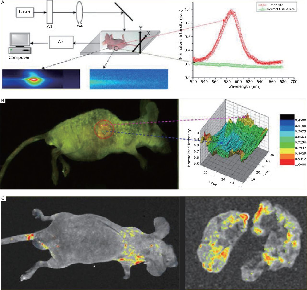
- 2
In vivo targeting and imaging of a lung metastasis model with QD-based nanotechnology. A: The imaging system for living animal models. B: In vivo targeted imaging of the subcutaneous tumor model and site-by-site spectra analysis of the tumor, which showed that the QD-labeled anti-a-fetoprotein monoclonal antibody probes per field were lower at the center than in the periphery of the tumor, indicating that tumor growth was not homogeneous and the peripheral site was more active. C: In vivo targeted imaging of liver cancer lung metastasis models. Reproduced with permission from [53].
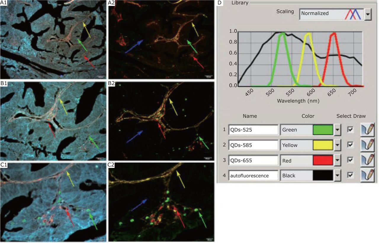
- 3
The establishment of multiplexed QD imaging and spectrum analysis. A1, B1, and C1: Infiltrating macrophages (Green arrows), type IV collagen (Yellow arrows), and neovessels (Red arrows) are labeled simultaneously in gastric cancer tissues with nanoprobes QDs-525, QDs-585, and QDs-655, respectively. A2, B2, and C2: Corresponding unmixed image of A1, B1, and C1 obtained by spectrum analysis with differentiable autofluorescence (Blue arrows). D: QD emission spectra and tissue autofluorescence data used for unmixed image. Magnification: ×100 (A1 and A2), ×200 (B1 and B2), and ×400 (C1 and C2); Scale bar: 100 mm (A1 and A2), 50 mm (B1 and B2), and 20 mm (C1 and C2). Reproduced with permission from [103].
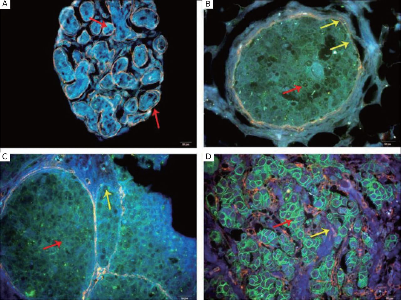
- 4
Double-color imaging was used in BC over different levels of HER2. A: Benign breast tumor, no HER2 expression, and intact ECM (Red arrow). B: BC with HER2 (+) (Red arrow), ECM becomes unsmooth and thin (Yellow arrows). C: HER2 (2+) and moderate green fluorescence (Red arrow). ECM becomes significantly degraded (Yellow arrow). D: HER2 (3+), strong green fluorescence (Red arrow) and complete ECM degradation (Yellow arrow). (Magnification: ×, scale bar =20 µm). Reproduced with permission from [69].


Tables
- 1
Comparison of the characteristics and applications between traditional organic fluorophores and QDs.
Property Traditional organic fluorophores[32–35] Quantum dots[29, 36–42] Chemical properties Chemical resistance is often poor Resistant to chemical degradation; sensitivity to pH determined by coatings Size scale Molecular, <0.5 nm Colloidal, 1.5 nm to 10 nm diameter Hydrodynamic radius Small, <0.6 nma Variable, 1.4 nm to 40 nmb Absorption spectra Discrete bands, FWHMc, 35 nmd to 80 nm to 100 nme Strong and broad Emission spectra Broad, red-tailed, and asymmetric, FWHM, 35 nm to 70 nm to100 nm Narrow, symmetric, FWHM, 30 nm to 90 nm Two-photon cross-section 10 GM to 500 GM (2,000 to 47,700) GMf Molar absorption coefficient (103 to 105) cm-1mol-1L (105 to 106) cm-1mol-1L Quantum yield Variable, 0.05 to 1.0 High, >20%g Fluorescence lifetime Short, <5 ns, mono-exponential decay Long, >10 ns, typically multi-exponential decay Solubility or dispersibility Control by substitution pattern Control via surface chemistry (ligands) Thermal stability Dependent on dye class; can be critical for NIR-wavelength dyes High; depends on shell or ligands Photostability Usually poor Excellent resistance to photobleaching; observation time of minutes to hours Bioconjugation labels Monovalent to multivalent labeling possible Scaffolds; Monovalent conjugation can be challenging; distribution of multivalences often encountered Applicability to single molecule analysis Moderate; limited by photobleaching Good; limited by blinking Spectral Multiplexing Possible Ideal for multi-color experiments; up to five colors demonstrated Multifunctionality Difficult and few Great potential Toxicity Variable, based on dye Related to the heavy metal a:Except for fluorescent proteins, GFP 4.6×2.4 nm cylindrical shape
b:Coating, ligand, and bioconjugate-dependent
c:FWHM, full width at half height of the maximum.
d:Dyes with resonant emission, such as fluoresceins, rhodamines and cyanines.
e:CT dyes.
f:Wavelength-dependent; GM: Goeppert–Mayer units
g:Ligand, coating and solvent-dependent
Probes Modality Size (nm) Application References 64Cu-DOTA to CdTe/ZnS (QD705) PET/optical ND Tumor vasculature imaging [100,122,123] 64Cu-DOTA to CdSe/ZnS (QD525, QD800) PET/optical ND Quantitative biodistribution in living mice [124] Gd-DOTA to CdSeTe/CdS/glutathione MRI/optical 7 to 10 Lymph node imaging of mouse [125] Iron oxide and CdSe/ZnS micelle MRI/optical 6.7 Simultaneous targeted drug delivery and dual-mode imaging of tumor tissues by near-infrared fluorescence and NMR spectroscopy [126] Resolve Al-Gd and CdSe/ZnS micelle MRI/optical 18 Tracing blood circulation in vivo [127] Gd-lipid in coating and CdSe/ZnS/silica MRI/optical 15 Tumor angiogenesis imaging [115] MnCdTeSe/CdS MRI/optical 4 to 50 Pancreatic cancer imaging [128]
In this issue


Jump to section
Related Articles
Cited By...
- No citing articles found.