

Abstract
OBJECTIVE To investigate the in vitro lethal effect of photodynamic therapy (PDT) using the photosensitizer hematoporphyrin on the human pancreatic cancer cell line Panc-1, the major influencing factors and the mechanisms of treatment.
METHODS Three factors—the time needed for photosensitizer and cell incubation, the photosensitizer concentration (PhoC) and the exposure dose (ExpD)—were examined with different levels of these factors. Optical density (OD) was used as a measure of CCK-8 in the experiment, and was converted to the rate of cell survival. The separate effect of each factor on the photodynamic action was studied, and the interactions were investigated. The effects of different incubation times and PhoC levels on the fluorescence intensity (FI) of the intracellular photosensitizer were determined, and the mechanisms of these factors leading to the therapeutic effects of PDT discussed.
RESULTS An increase in the photosensitizer and cell incubation time, an increase of PhoC, and enhancement of the ExpD, produced a corresponding decrease in the rate of Panc-1 cell survival after PDT (P < 0.05). PDT achieved its maximum lethal effects 16 h after starting the incubation, with a PhoC of 10 mg/L and an ExpD of 20 J/cm2; at these levels a synergistic interaction between PhoC and the ExpD occurred, decreasing the cell survival rate (P < 0.05). Neither simple administration of photosensitizer without ExpD (0 J/cm2) or illumination in the absence of PhoC (0 mg/L) affected the rate of cell survival (P > 0.05). With an increase of PhoC and lengthening of the incubation time, the FI of the intracellular photosensitizer accordingly increased (P < 0.05), and attained its maximum value at a PhoC of 10 mg/L and 36 h after the incubation. With an increase of PhoC, the FI of the photosensitizer, hematoporphyrin, in the solution increased progressively at first and then decreased (fluorescence quenching).
CONCLUSION PDT with the photosensitizer hematoporphyrin has clear lethal effects on the human pancreatic cancer cell line Panc-1, but the presence of a photosensitizer and laser irradiation by themselves do not have independent lethal effects. The three influencing factors—the time for photosensitizer and cell incubation, PhoC and ExpD—correlate positively with the PDT response, within certain limits. Beyond these limits, the PDT response does not significantly increase. The main mechanism of the PDT response lies in the effect of these factors on the level of the intracellular photosensitizer and the fluorescence quenching of the photosensitizer. A synergistic effect exists between PhoC and ExpD.
keywords
Introduction
The survival rate from pancreatic cancer is the lowest of all the common malignant tumors and surgery is possible in only about 10% of patients[1]. For those with non-operable pancreatic cancer, the available therapeutic regimens are chemotherapy, radiotherapy or combined therapy with both. The 1-year overall survival rate is < 10%[2], so a new therapy is urgently needed.
Photodynamic therapy (PDT) is a new method of tumor treatment which was developed in the 1980s. The principle of PDT is as follows: a photosensitizer can be selectively accumulated in the tumor, and then activated using a laser of specific wavelength, thus producing a singlet oxygen and oxygen radical, which can destroy the tumor tissues[3]. PDT has been mainly used to treat tumors on the body surface or inside the respiratory tract and digestive canal. However, over the past few years progress has been made in technologies such as ultrasonic endoscopy which will make it possible to use PDT to treat tumors in deep internal organs—for example, intractable diseases such as pancreatic cancer.
Studies on the response of pancreatic cancer to PDT have been mainly carried out in nude mice, and there are few reports from China. In this research, we used the most commonly seen human ductal pancreatic cancer cell line Panc-1 as the target of treatment, and the lethal effect of PDT on the in vitro cultured pancreatic cancer cells was systematically observed. Also, the main influencing factors and mechanisms were discussed.
Materials and Methods
Materials
The human pancreatic cancer cell line Panc-1 was bought from the American Type Culture Collection (ATCC), USA, and was frozen and stored in the medical research center of the Memorial Hospital. The photosensitizer Photosan, a freeze-dried injectable powder (hematoporphyrin, 145 mg/A), was purchased from Seehof Laboratory, Germany; the CCK-8 (Cell Counting Kit-8) from Japanese Dojindo Laboratories, Japan; RPMI 1640 culture medium and penicillin-streptomycin (P-S) from Gibco Co, USA; fresh fetal calf serum from Hangzhou Jinuo Biomedical Co, Hangzhou, China; the Biolitec PDT 630 semiconductor laser therapeutic apparatus from CeramOptec GmbH Co, Germany; the flow cytometer (FACSCalibur) from Becton Dickinson Co, USA; and the combined time-resolved steady-state fluorescence spectrometer (CTRSFS) from the Edinburgh Instrument Co, UK.
Methods
In vitro culture of the human pancreatic cancer cell line Panc-1
In vitro culture was carried out according to the methods of situ[4]. Panc-1 cells were cultured in RPMI-1640 complete medium containing penicillin 100 U/mL, streptomycin 100 μg/mL, and 10% fresh fetal calf serum, then transferred to an incubator with 5% CO2 and at 37°C. The cell density was (1–2) ×106/mL, and one passage was made every 3–4 days. The logarithmic growth phase was used for the experiments.
Preparation of the photosensitizer solution
The freeze-dried powder Photosan was weighed (145 mg/A) before each experiment with an electronic analytical balance, RPMI-1640 medium in the absence of fetal calf serum was used to prepare solutions of different concentrations, and stored at 4°C until needed, according to the method of Yang et al.[5] Storage and all processes were carried out in the absence of light.
Characteristics of the human pancreatic cancer cell line Panc-1 absorption of the photosensitizer Photosan
Cell absorption of the photosensitizer was reflected by the change in the level of available photosensitizer in the cells. The photosensitizer Photosan is a hematoporphyrin, and the porphyrins have a conjugated ring structure which is fluorescent[6], so based on the principle of PDT, the relative fluorescence intensity (RFI) in cells could be detected by flow cytometry, reflecting the level of available intracellular photosensitizer.
i) Determination of the fluorescence spectrum of photosensitizer Photosan
A “CTRSFS” was used to detect the activation of Photosan and the wavelength of the irradiation, thus setting the channel parameters of the flow cytometer.
ii) Determination of fluorescence intensity (FI) after incubation of intracellular photosensitizer of various concentrations, and Panc-1 cells
The following concentrations of photosensitizer were prepared: 0.5 mg/L, 1 mg/L, 2 mg/L, 3 mg/L, 4 mg/L, 6 mg/L, 8 mg/L, 10 mg/L, and 12 mg/L. A blank control containing no photosensitizer was also prepared. Triple wells were produced for each concentration. The logarithmic growth phase of the Panc-1 cells was obtained. After trypsinization, the cells were washed three times with complete medium and the cell density adjusted to 1 × 10⁵/mL. Cells were then inoculated onto a 12-well culture plate, 1 mL in each well, and cultured for 24 h to allow adherent growth of the cells. The cell culture fluid was replaced by photosensitizer solutions containing the abovementioned concentrations, 1 mL/well, and incubated for 8 h. The cells from each group were harvested, and flow cytometry used to determine the RFI.
iii) Determination of the FI of the intracellular photosensitizer at different points of time after incubation of the photosensitizer and Panc-1 cells
The preparation and inoculation of the Panc-1 cells was carried out as described above. The photosensitizer concentration (PhoC) was set at 4 mg/L, and cell incubation carried out for 1 h, 2 h, 4 h, 6 h, 8 h, 12 h, 16 h, 20 h, 24 h, 36 h, 48 h and 60 h. A blank control containing no photosensitizer (0 h) was also prepared. Triple wells were produced for each time period. A 12-well culture plate was set up for each point of time, and the cell culture fluid was replaced by photosensitizer at 4 mg/L, 1 mL/well, and incubated continuously. At the end of the study, the cells of each group were harvested, and flow cytometry used to determine the RFI.
iv) Fluorescence quenching
Phosphate-buffered saline (PBS) solutions containing Photosan at concentrations of 0.8 mg/L, 1 mg/L, 2 mg/L, 4 mg/L, 5 mg/L, 8 mg/L, 10 mg/L, 20 mg/L, 40 mg/L, 50 mg/L, 80 mg/L and 100 mg/L were prepared. The fluorescence emission spectrum of the photosensitizer was measured, FI at each site was observed, and an FI-concentration curve was drawn based on the emission wavelength determined in section (a).
PDT effect of the photosensitizer Photosan on Panc-1 cells and the factors which influence it
Effect of PhoC and exposure dose (ExpD) on PDT response
i) PDT
The PhoC was as follows: 0.5 mg/L, 1 mg/L, 2 mg/L, 3 mg/L, 4 mg/L, 6 mg/L, 8 mg/L, 10 mg/L, and 12 mg/L; a blank control group with no photosensitizer was also set up. The ExpDs used were as follows: 1 J/cm2, 5 J/cm2, 10 J/cm2, 15 J/cm2, 20 J/cm2, 25 J/cm2, and 30 J/cm2; a blank control group with no exposure was also set up. Triple wells were produced for each group. The density of Panc-1 cells was adjusted to 5 × 10⁴/mL. The cells were inoculated onto a 96-well plate, 100 μL each well, and cultured for 24 h to allow adherent growth of the cells. The cell culture fluid was replaced by the photosensitizer solution at the concentrations mentioned above, 100 μL/well, and cultured for 8 h in an incubator. The photosensitizer solution was replaced by complete medium without fetal calf serum, 100 μL/well, and laser irradiation was immediately administered, at a wavelength of 630 nm and a light-spot diameter of 5 cm. The ExpDs between 1 and 30 J/cm2 were applied to the cells at every PhoC. To avoid the impact of light scattering and reflection, laser irradiation was performed on the culture plate once only. After exposure, all 96-well plates were put into the incubator and incubated continuously for 24 h.
ii) CCK-8 assay of Panc-1 cell absorption after PDT
The principles were as follows: CCK-8 agents contain WST-8, which produces a water-soluble formazan dye upon bioreduction in the presence of an electron carrier, 1-methoxy PMS. The quantity of formazans generated correlates directly with the number of living cells, and therefore, this can be used to detect the absorption by flow cytometry, enabling the cytotoxicity to be determined[7]. The method was as follows: 10 μL CCK-8 was added to each of the 96-well plates, mixed well by shaking gently for 10 min, and the plates removed after incubation for 4 h. Microplate scanning spectrophotometry was used to detect absorption in the wells of each group, and the zero adjustment set by one blank control well in each plate. The detected wavelength was 450 nm, and the reference wavelength 630 nm.
(b) Effects of the time for photosensitizer and cell incubation on PDT response.
i) PDT
The groups were divided according to the time for photosensitizer and cell incubation: 2 h, 4 h, 8 h, 12 h, 16 h, 20 h, and 24 h. A blank control containing no photosensitizer (0 h) was also prepared. Triple wells were produced for each concentration. The PhoC was set as 4 mg/L, and ExpD as 5 J/cm2. The cells were inoculated onto the 96-well plates, the medium was replaced by the photosensitizer solution and PDT exposure was the same as described in section (a).
ii) CCK-8 assay of the Panc-1 cell absorption after PDT
See also section (a). The above-mentioned two experiments, i.e. the determination of FI of intracelular photosensitizer and the CCK-8 assay of the absorbance after PDT, were repeated three times. All operations were conducted in the absence of light.
Statistical analysis
Results are shown as mean ± standard deviation. One-factor analysis of variance was used for comparison of the mean of multiple samples, and factorial analysis of variance was used for samples with two factors and multiple levels. A least significant difference test or Student-Newman-Keuls test was conducted. SPSS version 17.0 was used for all computations. A value of P < 0.05 was used to show statistically significant differences.
Results
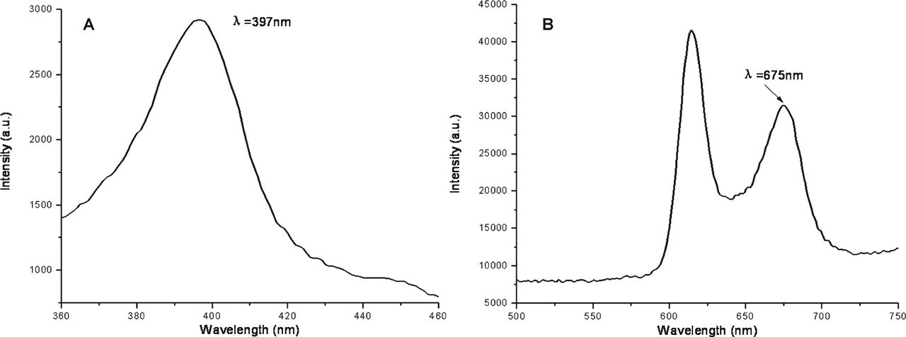
Photofluorogram of photosensitizer Photosan
Fig.1. Shows the fluorescence excitation spectrum (A) and emission spectra (B) of the detected Photosan. The excitation wavelength was 397 nm, and the emission wavelength 675 nm.

Fluorescence excitation spectrum (A) and emission spectra (B) of Photosan.
Characteristics of the human pancreatic cancer cell line Panc-1 absorption of the photosensitizer Photosan
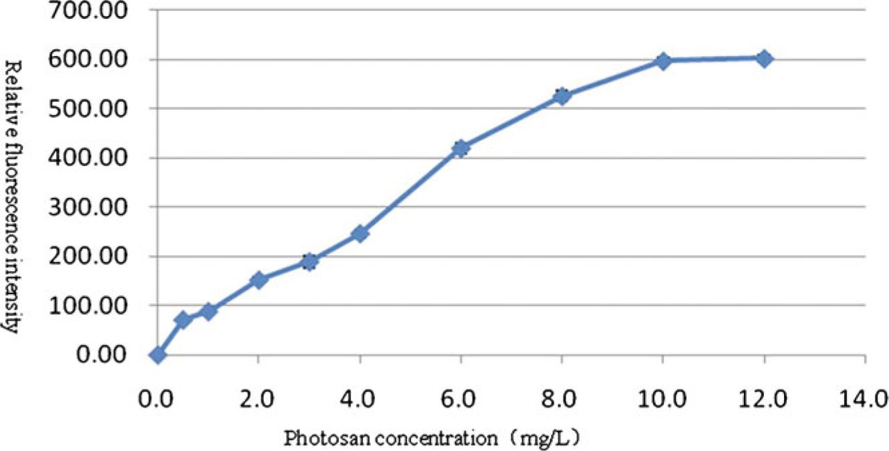
FI of the intracellular photosensitizer after incubation of the photosensitizer at various concentrations and with Panc-1 cells. Based on the results shown in Fig. 1, the selected channel parameter for flow cytometry was an excitation wavelength of 397 nm, and an emission wavelength of 675 nm. After incubation for 8 h, the intracellular RFI in the photosensitizer groups of various concentrations and in the blank control group were measured (Table 1).
Fluorescence intensity of the intracellular photosensitizer after its incubation at different concentrations and with Panc-1 cells.
It is clear, therefore, that with a constant incubation time the level of available intracellular photosensitizer increases as the PhoC increased. Thus, at low concentrations, the level of intracellular photosensitizer significantly increased as the concentration increased, and when the concentration was high (at ≥ 10 mg/L), the level of intracellular photosensitizer reached a plateau (Fig.2).

Fluorescence intensity of the intracellular photosensitizer after its incubation at different concentrations and with Panc-1 cells.
FI of the intracellular photosensitizer after incubation of the photosensitizer and Panc-1 cells for different lengths of time
The concentration of the photosensitizer was kept constant at 4 mg/L, and the intracellular RFI measured at different times of incubation (Table 2).
Fluorescence intensity of the intracellular photosensitizer at different points of time after incubation of the photosensitizer and Panc-1 cells.
It is clear that with a constant PhoC, the level of available intracellular photosensitizer increased as the incubation time increased. Thus, at the shorter incubation times the level of intracellular photosensitizer significantly increased as the incubation time increased, and when the incubation time was long (≥ 36 h), the level of intracellular photosensitizer reached a plateau (Fig.3).
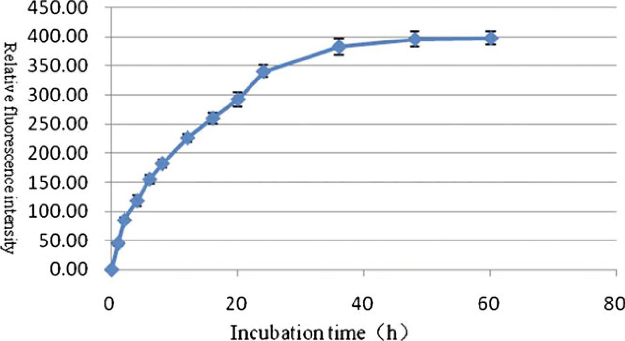
Fluorescence intensity of the intracellular photosensitizer after different lengths of incubation of photosensitizer and Panc-1 cells.
Fluorescence quenching of the photosensitizer in solution
Fig.4. shows the RFI of the emission wavelength of Photosan in solutions of various PhoCs. In the range 0.8–8 mg/L, the RFI increased with increasing PhoC. In contrast, when PhoC was > 8 mg/L, the RFI reduced with increasing PhoC. At a very high PhoC (100 mg/L), the RFI was even lower than that at 0.8 mg/l PhoC.

Fluorescence intensity curve of Photosan at different concentrations in phosphate-buffered saline solution (λEm 675 nm).
PDT effect of the photosensitizer Photosan on Panc-1 cells and its influencing factors
Effects of PhoC and ExpD on the PDT response
A CCK-8 assay showed that the optical density at 450 nm (OD450) in the zero-set 24 h after PDT was 0. Table 3 shows the OD450 value in the groups of various PhoC and the blank group.
OD450 value of Panc-1 cell lines after treatment with photosensitizer of different concentrations and exposure of various doses (mean ± SD).
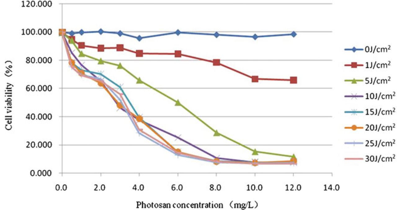
The rate of cell survival in each well can be calculated using the principle of the CCK-8 assay (Fig.5).

The cell survival rate of Panc-1 cells after treatment with photosensitizer of different concentrations and different exposure.
Cell-survival rate (%) − [(OD450 of the assay well – OD450 of the zero-set well)/(OD450 of the control well –OD450 of the zero-set well)] × 100%
The results showed that with an increase of PhoC and ExpD, the overall cell survival rate decreased after PDT. When PhoC was in the range 0.5–10 mg/L, the cell survival rate clearly decreased with the increase in concentration. When PhoC was >10 mg/L, no clear decrease in the cell survival rate was seen. When the ExpD was in the range 1–20 J/cm2, the cell survival rate fell as the dose increased. However, there were no significant differences between the groups with an ExpD of 10 J/cm2 and 15 J/cm2. The rate no longer decreased significantly at doses >20 J/cm2. In the absence of photosensitizer (0 mg/L), or illumination (0 J/cm2) cell survival rate did not change. Interaction occurred between the PhoC and ExpD (P<0.05). Statistical analysis using the interaction parameters was carried out[8]. A linear regression analysis was conducted for the variables “PhoC*ExpD” representing the interaction of the two influencing factors and OD450, and the regression coefficient β = −0.421 was obtained (P < 0.05), indicating a synergistic effect between the two factors.
Effect of the time for photosensitizer and cell incubation on PDT response
Twenty-four hours after PDT, the CCK-8 assay showed the value of OD450 in zero-set well was 0. Table 4 shows the OD450 value in the experimental and control groups.
The OD450 value at different points of time after incubation of photosensitizer and Panc-1 cells (mean ± SD).
Fig. 6 shows the rate of cell survival in all groups. Analysis showed that as the incubation time of the photosensitizer and cells increased, there was an overall decrease in the rate of cell survival after PDT. In addition, when the incubation time was in a range 0–16 h, cell survival dropped sharply as the incubation time was increased. For incubation times >16 h, the cell survival was stable with no further decrease occurring.

Cell survival rate at different times after incubation of photosensitizer and Panc-1 cells.
Discussion
Characteristics of the photosensitizer Photosan in human pancreatic cancer cell line Panc-1 absorption
The three main factors affecting PDT are the incubation time for photosensitizer and cells, photosensitizer concentration (PhoC) and exposure dose (ExpD)[9]. The effect of PDT is due to the available photosensitizer which is in the cells and which can be activated by the exposure, and thus, clearly, the two factors affecting PDF are the incubation time and the PhoC. To observe the effect of these two factors on the cells’ absorption of photosensitizer it is important to understand the time effect and the quantity effect separately so that a suitable PhoC and administration time can be selected.
The results showed that the absorption of the photosensitizer Photosan by the pancreatic cancer cell line Panc-1 increased as PhoC and incubation time increases. This increase gradually slowed down and plateaued as the two factors increased, reaching a maximum absorbance at a PhoC of 10 mg/L and an incubation time of 36 h.
We believe that this phenomenon may be explained as follows:
(a) Besides intake by the cell Panc-1 of photosensitizer, a process of “excretion” may also occur[10]. When the photosensitizer is first added and for some time after, the intake of the photosensitizer by the cells exceeds the excretion, thus showing an increased absorption. When the extracellular PhoC or the incubation time has increased to a certain point, the intake and excretion reach a dynamic equilibrium. At this time, the absorption has reached “saturation” and does not change.
(b) Another factor affecting the FI of the intracellular photosensitizer is fluorescence quenching. This is a physical or chemical process resulting from interaction between the fluorescent material and solvent molecules or in the molecules themselves owing to concentration self-quenching, which results in an attenuation or removal of FI[11]. Since Photosan is a molecule containing a large porphyrin ring, it is easy to produce stacking in the solution owing to a weak interaction between the rings, called a π-π accumulation. This may result in fluorescence quenching between molecules, which is particularly prominent at a high concentration[12]. Therefore, a Photosan solution of various concentrations was prepared, and the RFI was determined. Our results showed that with a gradual increase of Photosan concentration, FI is first enhanced and then attenuated. So, at a high PhoC and after a long incubation time, fluorescence quenching of the intracellular photosensitizer is one of the reasons why the FI no longer markedly increases.
(c) FI may be affected by complete mobilization of the receptors of the photosensitizer, which participate in cell conjugation and movement.
Our experiment suggests that with appropriate adjustment of the dose and length of administration of the photosensitizer, a curative effect of PDT in pancreatic cancer may be achieved
PDT response in the pancreatic cancer cell Panc-1 and the factors influencing it
The experimental results and analysis show that PDT had a clear lethal effect on the cell line Panc-1. Nevertheless, administration of the photosensitizer alone with no exposure (ExpD 0 J/cm2), or single exposure with no administration of photosensitizer (PhoC 0 mg/L), will not produce a PDT response, demonstrating that the presence of a photosensitizer and illumination at a specific wavelength are the essential factors needed for killing the cancer cells. This is in line with the basic characteristics of PDT[13]. This also suggests that the photosensitizer and the laser of relative wavelength do not have independent biological effects, and thus contact of one of these with normal tissue is safe.
The effects of PhoC and ExpD on PDT response were observed, and the values of the factors graded according to Yang et al.[5] and Song et al.[14] The results indicated that when these factors were doubled, the lethal effects were raised correspondingly. However, when the concentration of photosensitizer reached 10 mg/L, and the ExpD reached 15 J/cm2, the lethal effect did not significantly increase steadily. A “saturation” point in the PDT response was arrived at, owing to the cells reaching their maximum absorbance of the photosensitizer or to a saturation dose with the exposure in the cells. The laser FI might have resulted in an almost maximal photochemical reaction from the photosensitizer. Another possibility is that fluorescence quenching in the photosensitizer Photosan might have weakened the photodynamic action.
The results also showed that there was an interaction between PhoC and ExpD. To further ascertain the type of interaction and its effect on the results, a statistical analysis of the interaction coefficients was conducted. When the coefficient β≠0, this signifies some interaction between PhoC and ExpD, and a positive or negative value of β indicates, respectively, whether the interaction between the dependent variables is enhanced or weakened[8]. After calculation of the coefficients, the value of β was –0.421, showing that the interaction between PhoC and ExpD synergistically lowers the rate of cell survival. The reason for this phenomenon remains unclear, and no reports on this subject have been published. However, this phenomenon can still be used for economic guidance and to obtain a clinically high performance. For example, the curative effect can be increased by increasing the ExpD without adding more photosensitizer, and thus reduce the risk of side effects such as damage to the skin caused by a large dose of photosensitizer, and the adverse effect of fluorescence quenching. Increasing the ExpD has little effect on the cost, but increasing the PhoD will obviously increase the financial burden of patients.
A “time–effect ” relationship was also found when observing the effect of the length of incubation of photosensitizer and cells on the PDT response. Absorption of Photosan was seen from 1 h after the incubation until its lethal effect occurred, and was enhanced as the incubation time was increased. When the incubation time reached 36 h a plateau was reached and from that point onwards further extension of the incubation time did not significantly increase the lethal effect. Thus the incubation time affects the cell absorption of the photosensitizer, and the lethal effect of PDT.
Conflict of interest statement
No potential conflicts of interest were disclosed.
Acknowledgements
This work was supported by grants from Guangdong Provincial Natural Science Foundation (06021369) and Guangdong Medical Research Funds (B2006043).
- Received May 4, 2011.
- Accepted June 10, 2011.
- Copyright © 2011 by Tianjin Medical University Cancer Institute & Hospital and Springer
In this issue


Jump to section
Related Articles
Cited By...
- No citing articles found.