

Abstract
OBJECTIVE To investigate the correlations between the expression of thyroid transcription factor-1 (TTF-1) and apoptosis and angiogenesis in lung carcinomas.
METHODS A 829 microarray of the paraffin tissue chips was constructed, which contained 196 lung carcinomas, 10 normal lung tissues, and 1 muscular tissue. Terminal deoxynucleotidyl transferase mediated nick end labeling (TUNEL) and immunohistochemical SP method were used to detect apoptosis and expression of TTF-1 and CD34 in different types of lung carcinomas. A Leica Q500 MC image analysis system was used to measure and calculate TTF-1 positive unit (PU), apoptotic index (AI) and microvessel density (MVD).
RESULTS AI of lung small cell carcinoma and large cell carcinoma were smaller than those of lung adenocarcinoma and squamous cell carcinoma (P = 0.000). AI of lung carcinomas with lymph node metastases was smaller than that of those without (P = 0.039). AI of lung carcinomas in TNM stage I-IV was smaller than that in stage I (P = 0.008). The PU of the TTF-1 was negatively correlated with AI in small cell lung carcinoma (r = -0.752, P = 0.000). MVD of lung carcinomas without lymph node metastases was smaller than that of those with lymph node metastasis (P = 0.031). MVD of lung carcinomas in TNM stage I was smaller than that in stage I-IV (P = 0.040). The PU of TTF-1 was positively correlated with MVD in lung adenocarcinoma (r = 0.708, P = 0.000).
CONCLUSION There is a negative correlation between TTF-1 PU and AI in small cell lung carcinoma. TTF-1 PU and AI may be correlated with each other. There is a positive correlation between TTF-1 PU and MVD in lung adenocarcinoma. TTF-1 may induce the development of lung adenocarcinoma by inducing tumor angiogenesis.
keywords
Introduction
Thyroid Transcription Factor-1 (TTF-1) is an activating transcription factor of thyroid specific genes. TTF-1 expression differs in different types of lung carcinomas. It is higher in lung adenocarcinoma and small cell lung carcinoma and lower in squamous cell lung carcinoma and large cell lung carcinoma[1-3]. Malignant tumorigenesis is closely related to increased numbers of malignant cells, blockage of apoptosis and increased tumor angiogenesis[4]. Here we aim to investigate the correlations between TTF-1 and apoptosis and angiogenesis. We study the intensities of TTF-1 expression, levels of apoptosis and microvessel density in 196 lung carcinomas by immunohistochemistry, terminal-deoxynucleotidyl transferase mediated nick end labeling, and tissue microarray. We also employ quantitative analysis to investigate the correlations between TTF-1, apoptosis and angiogenesis.
Materials and Methods
Materials
A series of paraffin-embedded specimens were obtained from the Pathology Department, Nanfang Hospital, Guangzhou, China from January, 2003 to December, 2004. The patients whom the specimens were taken from were not treated with chemo or radiation therapies. The histological typing of lung carcinomas was conducted according to the third edition of Histological Typing of Lung Tumors, WHO/IASLC[5], in which included 69 adenocarcinomas (10 well-differentiated and 59 moderately to poorly differentiated), 84 squamous cell lung carcinomas (37 well-differentiated and 47 moderately to poorly differentiated), 27 small cell lung carcinomas and 16 large cell lung carcinomas. There were 98 cases of lung carcinomas with lymph node metastases (41 metastases from adenocarcinomas, 38 from squamous cell carcinomas, 13 from small cell lung carcinomas and 6 from large cell lung carcinomas). Seventy-nine lung carcinomas were of peripheral type and 117 were of central type. Eighty lung carcinomas were in stage I and 116 were in stages I~IV according to TNM (“tumor node metastasis” classification in the article). All paraffin-embedded tissue blocks were used as donor blocks.
Tissue microarray construction
For tissue microarray construction, a hematoxylin and eosin (H&E)-stained section was made from each donor block to define representative tumor regions. Tissue cylinders from muscle tissues (negative control, 5 cores), normal lung tissues (positive control of anti-TTF-1 antibody), and lung carcinomas (4 cores from each of the cases ranged from the cases with lymph node metastases to those without) were then punched from each donor tissue block and brought into a recipient paraffin block using a custom-made precision instrument (Beecher Instruments, MTA-1). A 25 × 33 matrix of 829 disks was constructed with 4 tissue cores in the first row to be an indication of the location[6-8].
Immunohistochemistry and Quantitative Analysis
Monoclonal antibodies to TTF-1 (Clone: 8G7G3/1, working solution) and CD34 (Clone: QBEnd/10, working solution), SP detection kit and DAB kit were products imported from the USA. Immunohistochemical staining was carried out on 3μm-thick sections in the constructed tissue microarray. Sections were deparaffinized regularly. Deparaffinized sections were first placed in plastic jars filled with citrate buffer (pH 6.0) and incubated for 10 min in an autoclave. The peroxidase reaction was performed using 3,3’-diaminobenzidine tetrahydrochloride (3,3’-diaminobenzidine) for 5 min.
Sections used for quantitative analysis were not counterstained. Suspicious staining was re-stained. PBS was used as a blank control. Muscle tissues served as negative controls and normal lung tissues as internal positive controls. A Leica Q500 MC image analysis system was used to detect the intensities of TTF-1 and CD34 staining. For TTF-1, according to the staining of the blank, positive and negative controls, the premise that there were no false positives or false negatives was confirmed. Those with dark-brown particles in the nuclei were regarded as positive expressions. Positive unit of each specimen was calculated[9-11]. For CD34, those with dark-brown particles in the cytoplasm of vascular endothelial cells were regarded as signs of expression. The parenchyma of the lung carcinomas was regarded as measuring areas[12]. Detection was done with a 40 × objective lens, microvascular areas were measured within 20 randomly selected fields in 4 randomly chosen representative cores of the tissue for each specimen. The averages of microvessel areas in the 20 fields selected were recorded as the total microvessel areas. Measuring areas in the 20 selected fields were measured respectively and the average was the total measuring areas. MVDs of 196 cases of lung carcinomas were calculated according to the following formula: MVD = total microvessel areas/total measuring areas.
Detection of apoptosis by TUNEL
Apoptosis detection kit (POD, MK1020) from BOSTER of Wuhan was used. TBS instead of TUNEL mixed solutions served as blank controls. Sections without TdT enzyme digestion served as negative controls and breast carcinoma as positive controls. The apoptosis index was calculated using the formula AI = (positive apoptotic cells/500 cells) × 100%. With a 40 × objective lens, the average number of positive cells within 10 detected fields was recorded as AI[13].
Statistical analysis
The SPSS 10.0 software package was used for statistical analysis with a cut-off of P < 0.05. TTF-1, CD34 and apoptosis in different groups of lung carcinomas were compared by ANOVA. Student’s t test was used to compare differences between clinicopathological parameters in different types of lung carcinomas. Spearman correlation analysis was used for the study of correlations between TTF-1 and AI, and TTF-1 and MVD.
Results
Immunohistochemistry and apoptosis results from the 829 disk tissue microarray
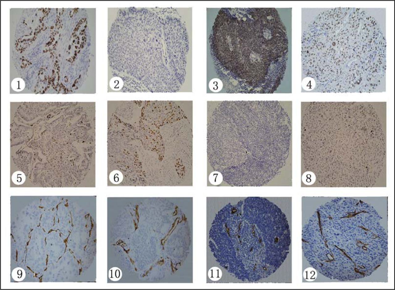
Results of immunohistochemistry and apoptosis within 829 disks of the tissue microarray are shown in Fig. 1.

Results of TTF-1 and CD34 Immunohistochemistry and Apoptosis in Different Types of Lung Carcinomas (Immunohistochemical SP Method, TUNEL, ×200 ). (1) TTF-1 Expression in Lung Adenocarcinoma; (2) TTF-1 Expression in Lung Squamous Cell Carcinoma; (3) TTF-1 Expression in Small Cell Lung Carcinoma; (4) TTF-1 Expression in Large Cell Lung Carcinoma; (5) Apoptosis in Lung Adenocarcinoma; (6) Apoptosis in Squamous Cell Lung Carcinoma; (7) Apoptosis in Small Cell Lung Carcinoma; (8) Apoptosis in Large Cell Lung Carcinoma; (9) CD34 Expression in Lung Adenocarcinoma; (10) CD34 Expression in Lung Squamous Cell Carcinoma; (11) CD34 Expression in Small Cell Lung Carcinoma; (12) CD34 Expression in Large Cell Lung Carcinoma.
Relationships between TTF-1 PU, AI and MVD and clinicopathological parameters of lung carcinomas
Relationships between TTF-1 PU, AI and MVD and patients’ gender, general types, differentiation, lymph node metastasis and TNM stages are shown in Table 1.
Relationships between TTF-1 PU, AI and MVD and clinicopathological parameters of lung carcinomas.
Correlations between TTF-1 PU and AI in different types of lung carcinomas
Correlations between TTF-1 PU and AI in lung adenocarcinoma, squamous cell lung carcinoma, small cell lung carcinoma and large cell lung carcinoma are shown in Table 2.
Comparisons between TTF-1 PU and AI in different types of lung carcinomas.
Correlations between TTF-1 PU and MVD in different types of lung carcinomas
Correlations between TTF-1 PU and MVD in lung adenocarcinoma, squamous cell lung carcinoma, small cell lung carcinoma and large cell lung carcinoma are shown in Table 3.
Comparisons between TTF-1 PU and MVD in different types of lung carcinomas.
Discussion
TTF-1 is a kind of transcription factor which plays a decisive role in the development and maintenance of the normal functions of the lung [14]. Studies have shown that TTF-1 expression differs in different types of lung carcinomas. It is higher in adenocarcinoma and small cell carcinoma and lower in squamous carcinoma and large cell carcinoma. Our previous study showed that TTF-1 missense and synonymous mutations were present in squamous lung carcinoma and mucinous lung adenocarcinoma. TTF-1 expression was negative in mutational lung carcinomas [15]. This explains why TTF-1 expression differs in different types of lung carcinomas at the molecular level.
The appearance of apoptotic cells in tissues is the result of gene interaction and comprehensive regulation. Loss of control of apoptotic regulating mechanisms is closely related to carcinogenesis. With the TUNEL, we found that AI differs in 196 lung carcinomas. AIs of small cell lung carcinoma and large cell lung carcinoma are significantly smaller than those of lung adenocarcinoma and squamous cell lung carcinoma. AI of lung carcinomas is related to lymph node metastases and TNM stages. It shows that apoptotic levels are decreased with increasing malignancy and metastatic potential of the tumor.
It has been shown in this study, by analyzing correlations between TTF-1 PU and AI in different types of lung carcinomas, that TTF-1 PU of small cell lung carcinoma is inversely correlated with AI. TTF-1 may promote the development of small cell lung carcinoma by suppressing apoptosis. The development and progression of neuroendocrinal lung carcinomas are closely related to apoptosis. Small cell lung carcinoma is a kind of neuroendocrinal tumor which abnormally secretes TTF-1 like secreting ectopic hormones[16]. Increased secretion of TTF-1 is one of the causes of decreased apoptosis in small cell lung carcinoma. TTF-1 PUs of lung adenocarcinoma, squamous cell lung carcinoma and large cell lung carcinoma are not correlated with their AIs. Therefore, it reveals that the expression of the TTF-1 in these lung cancers is not directly influenced by apoptosis.
Angiogenesis is the most decisive step in tumor growth, infiltration and metastasis. We detected the expression of CD34 in vascular endothelial cells of lung carcinomas and measured MVD. We found that MVD was similar in lung adenocarcinoma, squamous cell carcinoma, small cell carcinoma and large cell carcinoma, but no significant difference. MVD of lung carcinomas is related to lymph node metastases and TNM stages. It has shown that with the increasing metastatic abilities and malignancy, there is an increase in number of new blood vessels in lung carcinomas. With the progression of TNM stages and increased metastases, there is an increasing trend in MVD and TTF-1. It indicates that cancer cells with metastatic potential, which have higher expression of TTF-1, are more prone to promote angiogenesis. MVD and TTF-1 are not related to patients’ sex, general types and differentiation. We conclude that MVD and TTF-1 are not influenced by clinicopathological parameters.
It demonstrates in this study, by analyzing correlations between TTF-1 and MVD in different types of lung carcinomas, that TTF-1 PU of lung adenocarcinoma is positively correlated with MVD. TTF-1 PU and MVD are not correlated with each other in lung squamous cell carcinoma, small cell carcinoma and large cell carcinoma. Other studies have shown that as a nuclear transcription factor, TTF-1 may have secreting function and secrete activating factors which promote and induce angiogenesis[16]. Our studies show that TTF-1 expression is interactively related to tumor angiogenesis. Determining if TTF-1 functions as an activating factor of angiogenesis in lung adenocarcinoma and participates in growth and formation of microvessels in tumors by regulating proliferation of vascular endothelial cells, requires further experimental exploration.
Footnotes
This work was supported by grants from the National Natural Sciences Foundation of China (No.30271462); the Science and Technology Planning Project of Guangdong Province, China (No.2KM04501S); the Principal Science and Technology Project of Guangzhou City (No.2003Z2-E0061, E0062).
- Revision received March 10, 2008.
- Accepted January 21, 2009.
- Copyright © 2009 by Tianjin Medical University Cancer Institute & Hospital and Springer
In this issue


Jump to section
Related Articles
Cited By...
- No citing articles found.