

Article Figures & Data
Figures
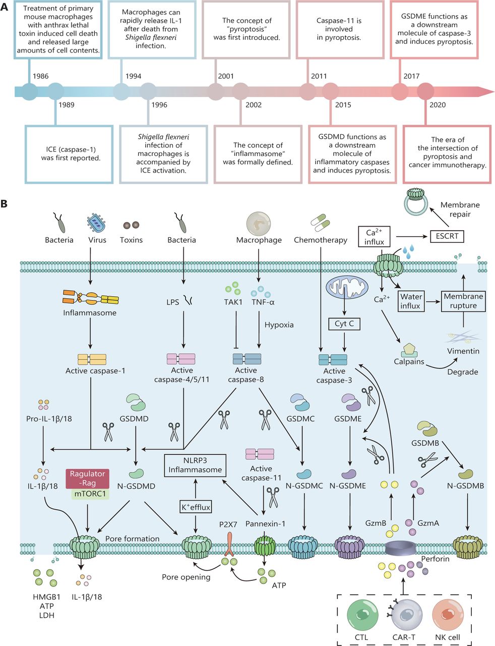
- Figure 1
The timeline and molecular mechanisms of pyroptosis. (A) Critical events in pyroptosis research. (B) Molecular mechanisms of pyroptosis. In the canonical pathway, upon stimulation with bacteria, viruses, or toxins, pattern recognition receptors on the cell surface activate downstream signaling pathways. Activated caspase-1 specifically cleaves gasdermin D (GSDMD) to produce GSDMD-N termini. The cleaved GSDMD-N fragment disrupts cell membrane integrity and induces cellular pyroptosis. In addition, activated caspase-1 promotes the production of mature IL-1β and IL-18, which induce an inflammatory response. Caspases-4/5/11 mainly mediate the noncanonical pathway of pyroptosis. Upon stimulation with bacterial lipopolysaccharide, activated caspases-4/5/11 specifically cleave GSDMD, leading to the formation of GSDMD pores that cause K+ efflux, thereby activating the NLRP3-caspase-1 signaling pathway and amplifying the inflammatory response. In addition, the formation of GSDMD pores is accompanied by Ca2+ efflux, which activates the calpain mechanism that promotes membrane rupture and the endosomal sorting complex required for the transport mechanism that promotes membrane repair. Various chemotherapeutic agents induce tumor cell pyroptosis by inducing the caspase-3-GSDME axis. Inhibition of TGF-β-activated kinase 1 (TAK1) induces pyroptosis in mouse macrophages through the caspase-8-GSDMD pathway. In addition, during hypoxic conditions, TNF-α produced by macrophages induces cancer cell pyroptosis using the caspase-8-GSDMC pathway.
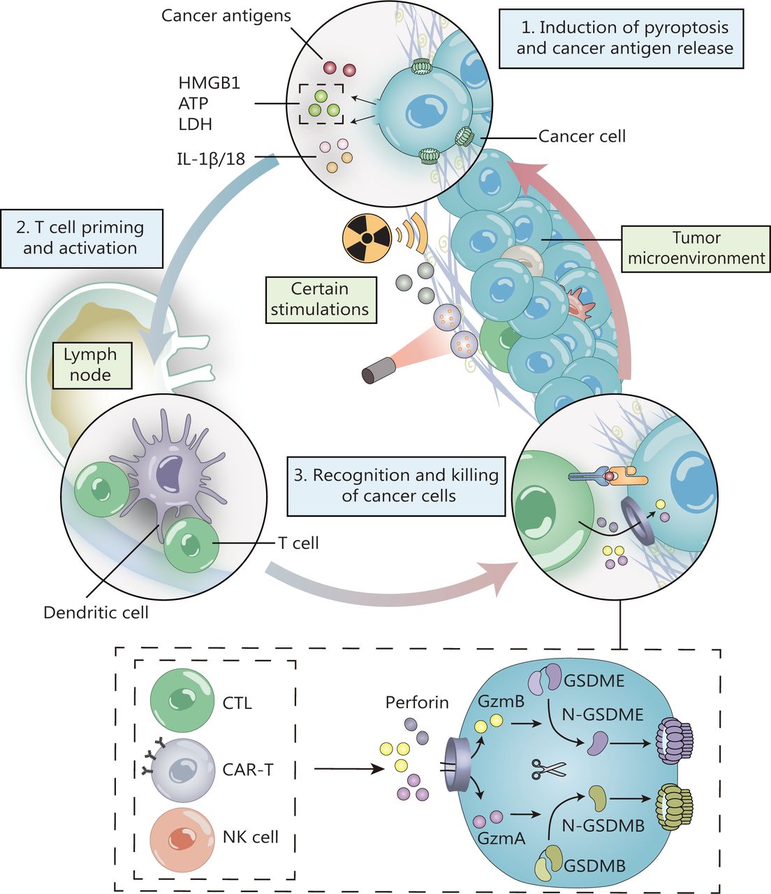
- Figure 2
The positive feedback loop of pyroptosis and anti-tumor immunity. Pyroptotic cells release cancer antigens and large amounts of inflammatory substances. These antigens are presented by dendritic cells and induce activation of T cells. Activated NK cells and cytotoxic T lymphocytes secrete granzyme A (GzmA) and B (GzmB). GzmA directly cleaves GSDMB, while GzmB cleaves caspase-3 and GSDME. The cleaved GSDMB-N and GSDME-N fragments induce cancer cell pyroptosis and enhance antitumor immunity, creating a positive feedback loop of pyroptosis and anti-tumor immunity.
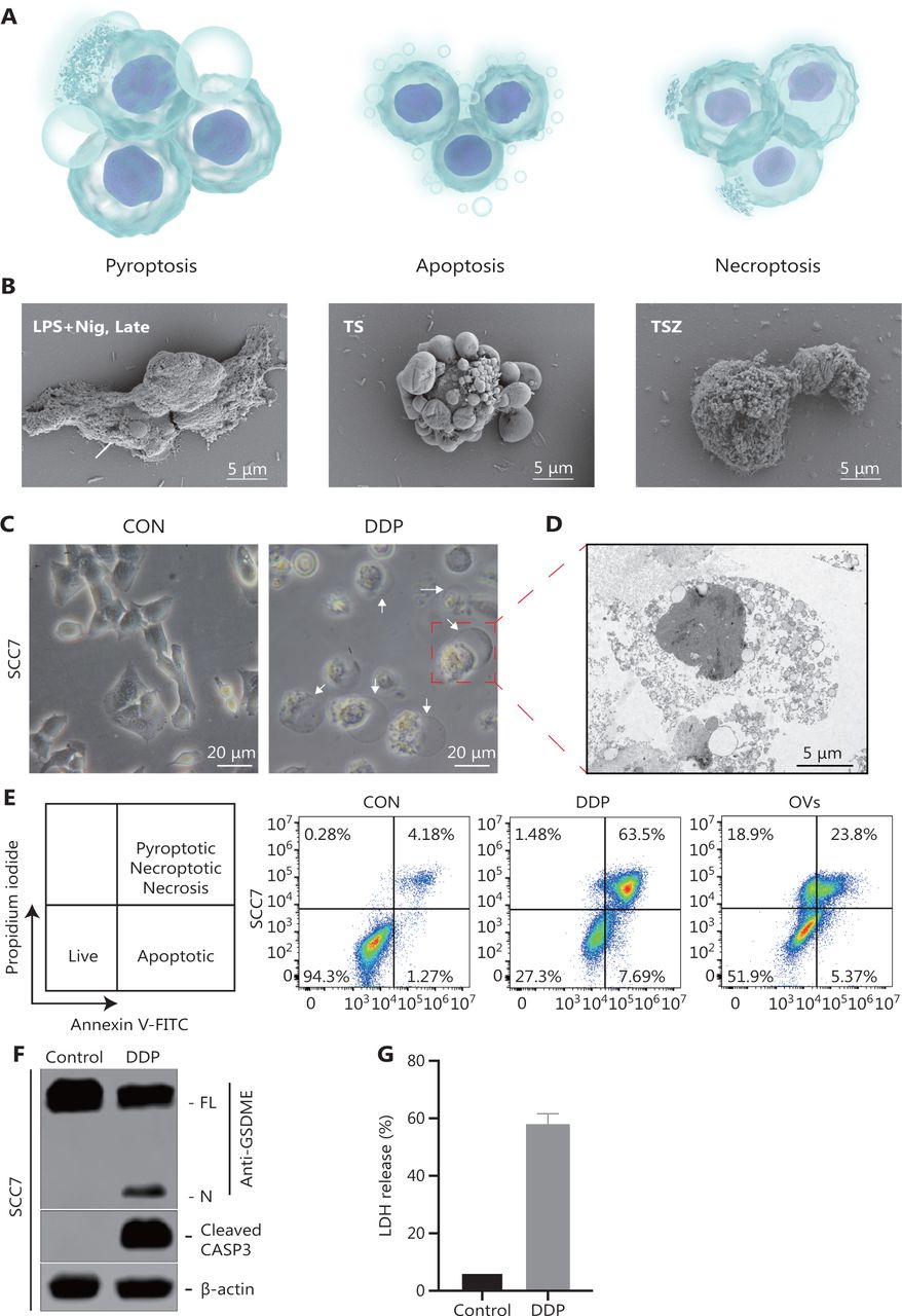
- Figure 3
Representative results of pyroptosis detection. (A-B) Cellular morphological features during pyroptosis, apoptosis, and necroptosis. (A) Schematic diagram showing the morphology of cells undergoing pyroptosis, apoptosis, and necroptosis. (B) Scanning electron microscopy of SCC7 cells exposed to different treatments, to distinguish different forms of cell death. The arrow points to the bubbling of pyroptotic cells. (C) Representative microscopic images of SCC7 cells after cisplatin (DDP) treatment. White arrowheads indicate characteristic ballooning in cell membranes. (D) Representative transmission electron microscopy images of SCC7 cells treated with DDP. (E) Flow cytometry analysis of propidium iodide (PI)- and Annexin V-stained cells. SCC7 cells were treated with DDP and oncolytic viruses, respectively, for 24 h. Annexin V−/PI− represents live cells, Annexin V+/PI− represents apoptotic cells, and Annexin V+/PI+ denotes pyroptotic or necroptotic cells. (F) Western blot analysis of pyroptotic cell death markers in SCC7 cells treated with DDP. (G) Cytotoxicity of SCC7 measured by detecting lactate dehydrogenase release in culture supernatants. Nig, nigericin; TS, TNF + SMAC mimetic; TSZ, TNF + SMAC mimetic + caspase inhibitor z-VAD.
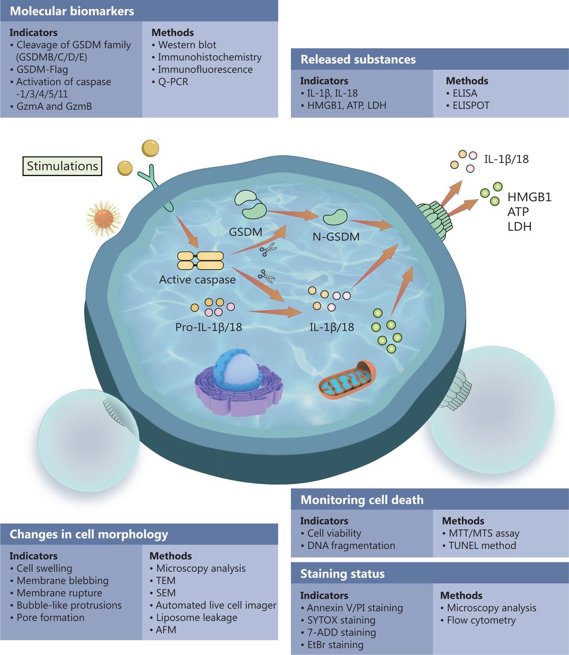
- Figure 4
Methods for monitoring pyroptosis. Pyroptosis is a multistep process. In practical applications, a combination of assays is often used to monitor the occurrence of pyroptosis and accurately determine pyroptosis results of experimental cells. GSDM, gasdermin; GzmA, granzyme A; GzmB, granzyme B; TEM, transmission electron microscopy; SEM, scanning electron microscope; AFM, atomic force microscopy.

Tables
Pyroptosis Apoptosis Necroptosis Character Active PCD Active PCD Passive PCD Inflammation Yes No Yes Morphology of cell membrane Cell swelling, membrane rupture, bubble-like protrusions Cell shrinkage, intact membrane, many vesicles of various sizes Cell rounding and swelling, membrane rupture Membrane blebbing Yes Yes No Membrane integrity No Yes No DNA damage Yes Yes Yes Chromatin condensation Yes Yes No Intact nucleus Yes No (fragmented) Yes Organelle morphology Deformation Intact Swelling Special constructions Pyroptotic bodies Apoptotic bodies No Release of intracellular contents DAMPs, inflammatory molecules No DAMPs, inflammatory molecules Associated molecules Initiation: Caspase-1, 3, 4, 5, 11.
Execution: GSDMD, GSDMEInitiation: Caspase-8, 9, 10.
Execution: Caspase-3, 6, 7
Pro-apoptotic members: Bax, Bak, Bok.
Anti-apoptotic members: Bcl-2, Bcl-xl, Mcl-1.Initiation: RIPK1/RIPK3.
Execution: MLKL.
Inhibitory members: Caspase-87-AAD staining Yes No Yes EtBr staining Yes No Yes PI staining Yes No Yes PS exposure Yes Yes Yes Annexin V staining Yes Yes Yes TUNEL staining Yes Yes Yes PCD, programmed cell death; GSDM, gasdermin; EtBr, ethidium bromide; PI, propidium iodide; PS, phosphatidylserine; TUNEL, TdT-mediated dUTP nick end labeling.
Indicators Method Reference Changes in cell morphology Cell swelling, membrane blebbing and rupture, bubble-like protrusions Microscopy analysis 23,27,48 TEM 64,65 SEM 21 Automated live cell imager 66 GSDM-mediated pore formation liposome leakage method 67,68 AFM 69,70 Monitoring cell death Cell viability MTT/MTS assay 71,72 DNA fragmentation TUNEL method 73,74 Staining status Annexin V/PI staining, SYTOX/7-ADD/EtBr/TO-PRO3 staining Microscopy analysis, Flow cytometry 23,25,53 Molecular biomarkers Cleavage of GSDM family (GSDMB/C/D/E) Western blot
Immunohistochemistry
Immunofluorescence
Q-PCR23,51,53,75,76 GSDM-Flag Activation of Caspase-1/3/4/5/11 GzmA and GzmB Released substances: IL-1β, IL-18, HMGB1, ATP, LDH ELISA, ELISPOT 27,72,77 Other methods The dynamic process of pyroptosis in vivo Two-photon imaging technology 78 TEM, transmission electron microscopy; SEM, scanning electron microscope; GSDM, gasdermin; AFM, atomic force microscopy; GzmA, granzyme A; GzmB, granzyme B.
Pyroptosis mechanism Detection Reference Caspase-3/GSDME Cell swelling with large bubbles 23 Cleavage of GSDME GSDME-mediated pore formation (liposome leakage method) ATP cell viability LDH release PI/FITC staining GZMB/(Caspase-3)/GSDME The changes in cell morphology 53 Cleavage of GSDME LDH, HMGB1 release SYTOX green uptake The use of zDEVD-fmk or zVAD-fmk to inhibit apoptosis and caspase-3-mediated pyroptosis GZMA/GSDMB The changes in cell morphology 51 Cleavage of GSDMB Edman sequencing of the N termini to identify the cleavage sites ATP–based cell viability LDH release PI/FITC staining Hypoxia/TNF-α/GSDMC Cell swelling with large bubbles 50 Cleavage of GSDMC Liposome leakage method Cell death determined by LDH release SYTOX green staining
In this issue


Jump to section
Related Articles
Cited By...
- No citing articles found.