

Article Figures & Data
Figures
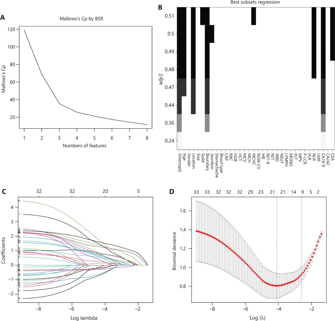
- Figure 1
Screening of model variables. Variables were screened with Best Subsets Regression (BSR) (A, B; AIC = 151.3), Lasso regression (C, D; AIC = 152.27), and univariate regression and multiple logistic regression (AIC = 144.02).
- Figure 2
(A) Multivariate analysis revealing that increased CA19-9, increased CEA, increased NLR, increased MCHC, non-O blood group, age > 55 years, cysts located in the head or neck of the pancreas, and an unclear cyst imaging border were independent predictors of PCN malignancy. (B) Nomogram for estimating the risk of malignancy in PCN. The nomogram is used to find the position of each variable on the corresponding axis, a vertical line is drawn to the points axis to obtain the value of the corresponding variable, the points are added for all variables, and then a line is drawn from the total points axis to determine the probability of a malignant tumor at the lower part of the nomogram.
- Figure 3
ROC analysis of the malignancy prediction ability of the sum of scores. (A) Internal validation of the nomogram. (B) External validation of the nomogram. (C, D) Nomogram calibration in the model group (C) and the validation group (D). The horizontal axis indicates the risk of malignant PCN predicted by the nomogram, and the vertical axis is the probability of malignant PCN for a random sample of 1000 actual observations. The line from the lower left to the upper right corner of the plot region represents the reference line of the ideal prediction.
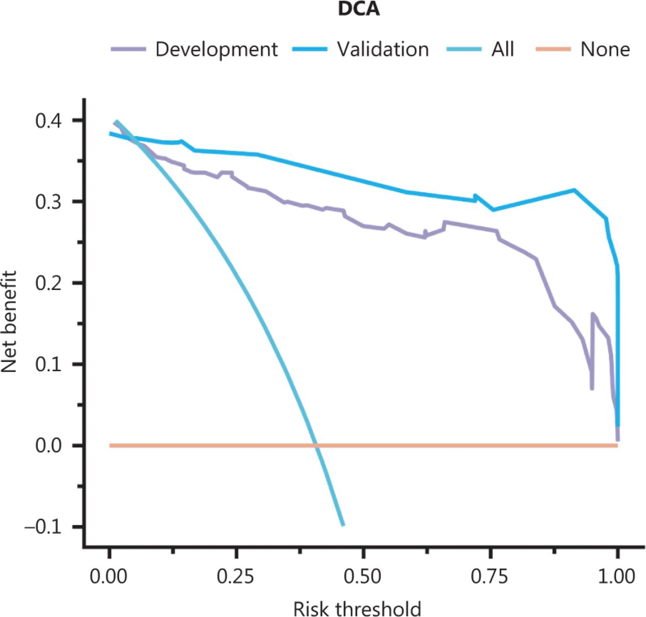
- Figure 4
DCA curves for assessing the net benefit of model application in the model development cohort and validation cohort.


Tables
- Table 1
Univariate logistic regression analysis based on preoperative data in the model cohort
Variable (%) Level Overall (%) Benign (%) Malignant (%) OR (95% CI) P n – 204 121 83 – – Age (years) ≤ 55 87 (42.6) 69 (57.0) 18 (21.7) 1.42 (1.25–1.61) < 0.001* > 55 117 (57.4) 52 (43.0) 65 (78.3) – – Gender Female 122 (59.8) 84 (69.4) 38 (45.8) 1.27 (1.11–1.45) 0.001* Male 82 (40.2) 37 (30.6) 45 (54.2) – – Location Body/tail 111 (54.4) 84 (69.4) 27 (32.5) 1.43 (1.26–1.63) < 0.001* Head/neck 93 (45.6) 37 (30.6) 56 (67.5) – – Size (cm) ≤ 2.75 158 (77.5) 97 (80.2) 61 (73.5) 1.1 (0.93–1.29) 0.265 > 2.75 46 (22.5) 24 (19.8) 22 (26.5) – – Solid No 79 (38.7) 62 (51.2) 17 (20.5) 1.37 (1.2–1.56) < 0.001* Yes 125 (61.3) 59 (48.8) 66 (79.5) – – Boundary Clear 100 (49.0) 83 (68.6) 17 (20.5) 1.59 (1.41–1.79) < 0.001* Blurred 104 (51.0) 38 (31.4) 66 (79.5) – – Jaundice No 187 (91.7) 120 (99.2) 67 (80.7) 1.79 (1.42–2.26) < 0.001* Yes 17 (8.3) 1 (0.8) 16 (19.3) – – Stomachache No 109 (53.4) 75 (62.0) 34 (41.0) 1.23 (1.07–1.4) 0.003* Yes 95 (46.6) 46 (38.0) 49 (59.0) – – Blood type Type-O 51 (25.0) 35 (28.9) 16 (19.3) 1.13 (0.97–1.32) 0.119 Non-O 153 (75.0) 86 (71.1) 67 (80.7) – – CRP (mg/L) ≤ 1.29 45 (22.1) 11 (13.3) 34 (28.1) 1.23 (1.05–1.45) 0.012* > 1.29 159 (77.9) 72 (86.7) 87 (71.9) – – RBC (×1012/L) ≤ 4.3 71 (34.8) 37 (30.6) 34 (41.0) 0.9 (0.78–1.03) 0.127 > 4.3 133 (65.2) 84 (69.4) 49 (59.0) – – HGB (g/L) ≤ 137.5 109 (53.4) 69 (57.0) 40 (48.2) 1.09 (0.95–1.25) 0.216 > 137.5 95 (46.6) 52 (43.0) 43 (51.8) – – HCT (%) ≤ 36.55 32 (15.7) 14 (11.6) 18 (21.7) 0.83 (0.69–1.00) 0.051 > 36.55 172 (84.3) 107 (88.4) 65 (78.3) – – MCV (fl) ≤ 90.65 93 (45.6) 60 (49.6) 33 (39.8) 1.1 (0.96–1.26) 0.168 > 90.65 111 (54.4) 61 (50.4) 50 (60.2) – – MCH (pg) ≤ 30.75 124 (60.8) 79 (65.3) 45 (54.2) 1.12 (0.97–1.28) 0.113 > 30.75 80 (39.2) 42 (34.7) 38 (45.8) – – MCHC (g/L) ≤ 341.5 174 (85.3) 109 (90.1) 65 (78.3) 1.25 (1.04–1.51) 0.020* > 341.5 30 (14.7) 12 (9.9) 18 (21.7) – – RDW.CV (%) ≤ 13.4 164 (80.4) 107 (88.4) 57 (68.7) 1.35 (1.15–1.6) < 0.001* > 13.4 40 (19.6) 14 (11.6) 26 (31.3) – – HR ≤ 2.875 43 (21.1) 16 (13.2) 27 (32.5) 0.76 (0.64–0.89) 0.001* > 2.875 161 (78.9) 105 (86.8) 56 (67.5) – – RETR (%) ≤ 0.985 24 (11.8) 11 (9.1) 13 (15.7) 0.86 (0.7–1.06) 0.154 > 0.985 180 (88.2) 110 (90.9) 70 (84.3) – – RET (×109/L) ≤ 82.4 158 (77.5) 90 (74.4) 68 (81.9) 0.90 (0.77–1.06) 0.207 > 82.4 46 (22.5) 31 (25.6) 15 (18.1) – – WBC (×109/L) ≤ 6.16 124 (60.8) 81 (66.9) 43 (51.8) 1.17 (1.02–1.34) 0.030* > 6.16 80 (39.2) 40 (33.1) 40 (48.2) – – NEUT (×109/L) ≤ 2.6 83 (40.7) 58 (47.9) 25 (30.1) 1.19 (1.04–1.37) 0.011* > 2.6 121 (59.3) 63 (52.1) 58 (69.9) – – LYMPH (×109/L) ≤ 1.905 132 (64.7) 71 (58.7) 61 (73.5) 0.86 (0.74–0.98) 0.030* > 1.905 72 (35.3) 50 (41.3) 22 (26.5) – – MONO (×109/L) ≤ 0.535 119 (58.3) 79 (65.3) 40 (48.2) 1.19 (1.03–1.36) 0.015* > 0.535 85 (41.7) 42 (34.7) 43 (51.8) – – PLT (×109/L) ≤ 207.5 68 (33.3) 34 (28.1) 34 (41.0) 0.87 (0.75–1.00) 0.056 > 207.5 136 (66.7) 87 (71.9) 49 (59.0) – – MPV (fl) ≤ 11.25 150 (73.5) 99 (81.8) 51 (61.4) 1.29 (1.11–1.5) 0.001* > 11.25 54 (26.5) 22 (18.2) 32 (38.6) – – P-LCR (%) ≤ 37.85 164 (80.4) 107 (88.4) 57 (68.7) 1.35 (1.15–1.6) < 0.001* > 37.85 40 (19.6) 14 (11.6) 26 (31.3) – – PLR ≤ 196.47 23 (11.3) 10 (8.3) 13 (15.7) 0.84 (0.68–1.03) 0.102 > 196.47 181 (88.7) 111 (91.7) 70 (84.3) – – NLR ≤ 1.79 92 (45.1) 67 (55.4) 25 (30.1) 1.28 (1.12–1.46) < 0.001* > 1.79 112 (54.9) 54 (44.6) 58 (69.9) – – LMR ≤ 3.64 61 (29.9) 22 (18.2) 39 (47.0) 0.72 (0.62–0.83) < 0.001* > 3.64 143 (70.1) 99 (81.8) 44 (53.0) – – CA19-9 (U/mL) − 146 (71.6) 109 (90.1) 37 (44.6) 1.72 (1.51–1.95) < 0.001* + 58 (28.4) 12 (9.9) 46 (55.4) – – CA242 (U/mL) − 170 (83.3) 117 (96.7) 53 (63.9) 1.77 (1.5–2.08) < 0.001* + 34 (16.7) 4 (3.3) 30 (36.1) – – CEA (μg/L) − 184 (90.2) 120 (99.2) 64 (77.1) 1.83 (1.48–2.26) < 0.001* + 20 (9.8) 1 (0.8) 19 (22.9) – – *P < 0.05 was considered statistically significant. CRP, C-reactive protein; RBC, red blood cell; HGB, hemoglobin; HCT, red blood cell specific volume; MCV, mean corpuscular volume; MCH, mean corpuscular hemoglobin; MCHC, mean corpuscular hemoglobin concentration; RDW.CV, red blood cell volume distribution width; HR, hemoglobin-red blood cell distribution width ratio; RETR, reticulocyte ratio; RET, reticulocyte; WBC, peripheral white blood cells; NEUT, neutrophils; LYMPH, lymphocyte; MONO, monocyte; PLT, platelet; MPV, mean platelet volume; P-LCR, platelet-larger cell ratio; PLR, lymphocyte-monocyte ratio; NLR, neutrophil-lymphocyte ratio; LMR, lymphocyte-monocyte ratio; CA19-9, carbohydrate antigen 19-9; CA242, carbohydrate chain antigen 242; CEA, carcinoembryonic antigen; OR, odds ratio; CI, confidence interval; +, CA199 > 37 U/ml, CA242 > 20 U/ml, CEA > 5 μg/L.
Variable Level Overall (%) Development cohort Validation cohort P No. patients 290 204 86 Age (years) ≤ 55 117 (40.3) 87 30 0.218 > 55 173 (59.7) 117 56 Location Body/tail 164 (56.6) 111 53 0.258 Head/neck 126 (43.4) 93 33 Boundary Clear 144 (49.7) 100 44 0.739 Blurred 146 (50.3) 104 42 Blood type Type-O 79 (27.2) 51 28 0.187 Non-O 211 (72.8) 153 58 MCHC (g/L) ≤ 341.5 241 (83.1) 174 67 0.125 > 341.5 49 (16.9) 30 19 NLR ≤ 1.79 132 (45.5) 92 40 0.825 > 1.79 158 (54.5) 112 46 CA19-9 (U/mL) ≤ 37 209 (72.1) 146 63 0.770 > 37 81 (27.9) 58 23 CEA (μg/L) ≤ 5 256 (88.3) 184 72 0.117 > 5 34 (11.7) 20 14 Imaging and tumor marker model Nomogram risk model Total Reclassified as higher risk (%) Reclassified as lower risk (%) Risk < 0.5 ≥ 0.5 < 0.5 No. of patients 117 33 150 33 (22%) NA No. of benign 105 7 112 7 (6.25%) NA No. of malignant 12 26 38 26 (68.4%) NA ≥ 0.5 No. of patients 4 50 54 NA 4 (7.4%) No. of benign 3 6 9 NA 3 (33.3%) No. of malignant 1 44 45 NA 1 (2.2%) Total No. of patients 121 83 204 33 (22%) 4 (7.4%) No. of benign 108 13 121 7 (6.25%) 3 (33.3%) No. of malignant 13 70 83 26 (68.4%) 1 (2.2%) NRI = 26.8%; P < 0.001.
IDI = 0.2708; P < 0.001.
In this issue


Jump to section
Related Articles
Cited By...
- No citing articles found.