

Article Figures & Data
Figures
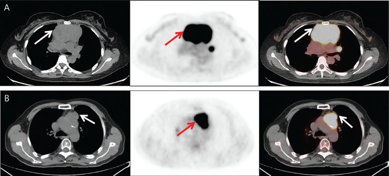
- Figure 1
Axial CT, PET, and fusion images (from left to right columns) of examples of primary mediastinal lymphomas (arrows) and thymic epithelial tumors (arrows). Panel A: 43-year-old female patient with diffuse large B-cell lymphoma (CT value = 4–38 Hu; SUVmax = 23.8 g/mL, MTV = 218.0 cm3, and TLG = 2774.9 g/mL cm3); panel B: 70-year-old male patient with squamous cell carcinoma (CT value = 39 Hu with scattered calcifications; SUVmax = 18.7 g/mL, MTV = 36.28 cm3, and TLG = 428.39 g/mL cm3).
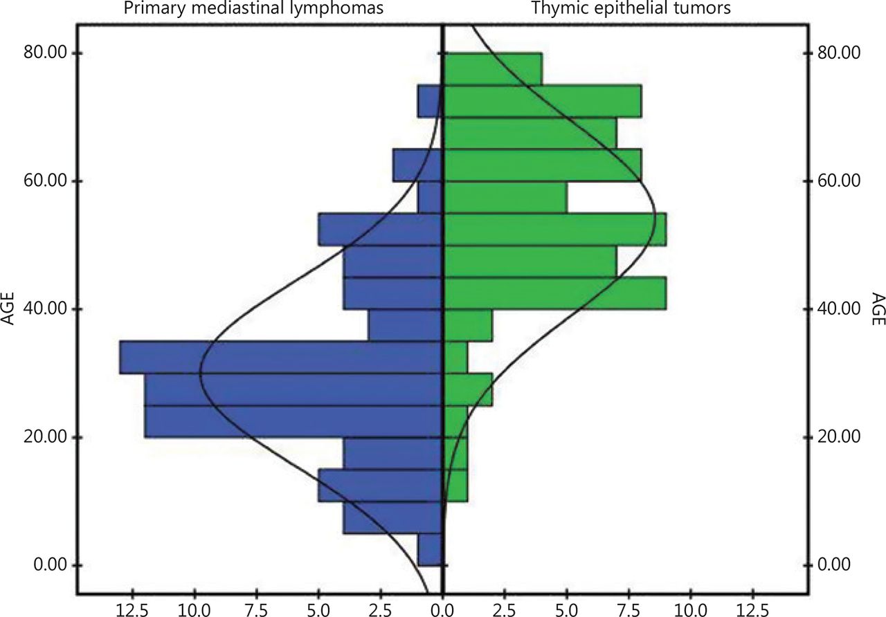
- Figure 2
Histograms of age distribution of the patients in this study, showing that most patients with primary mediastinal lymphomas (left panel) were < 40 years of age (76.1%), whereas most patients with thymic epithelial tumors (right panel) were > 40 years of age (87.7%).
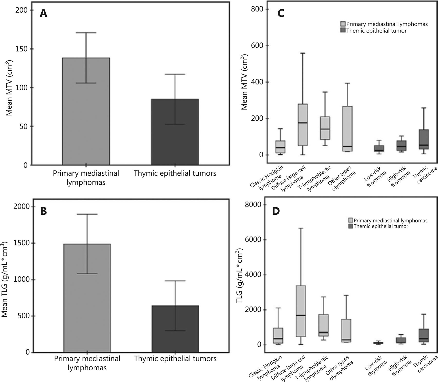
- Figure 3
The mean metabolic tumor volume (A) and total lesion glycolysis (B) in the patients with primary mediastinal lymphomas and thymic epithelial tumors, and a separate comparison for each subgroup of primary mediastinal lymphoma (C) and thymic epithelial tumors (D).
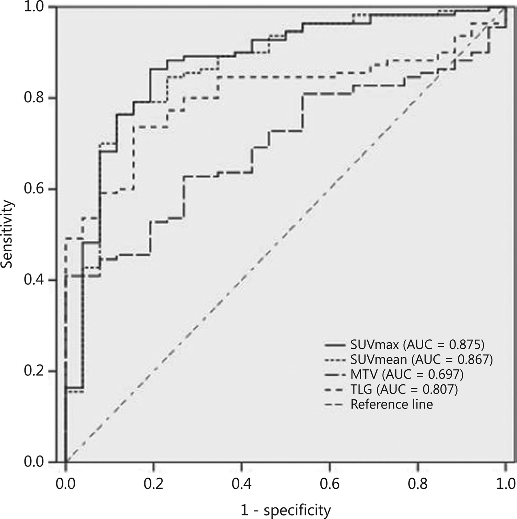
- Figure 4
ROC curve and area under the curve of the maximum standard uptake value (SUVmax), mean standard uptake value (SUVmean), metabolic tumor volume (MTV), and total lesion glycolysis (TLG) in differentiating patients with malignant tumors from patients with benign tumors in the pre-vascular compartment of the mediastinum.
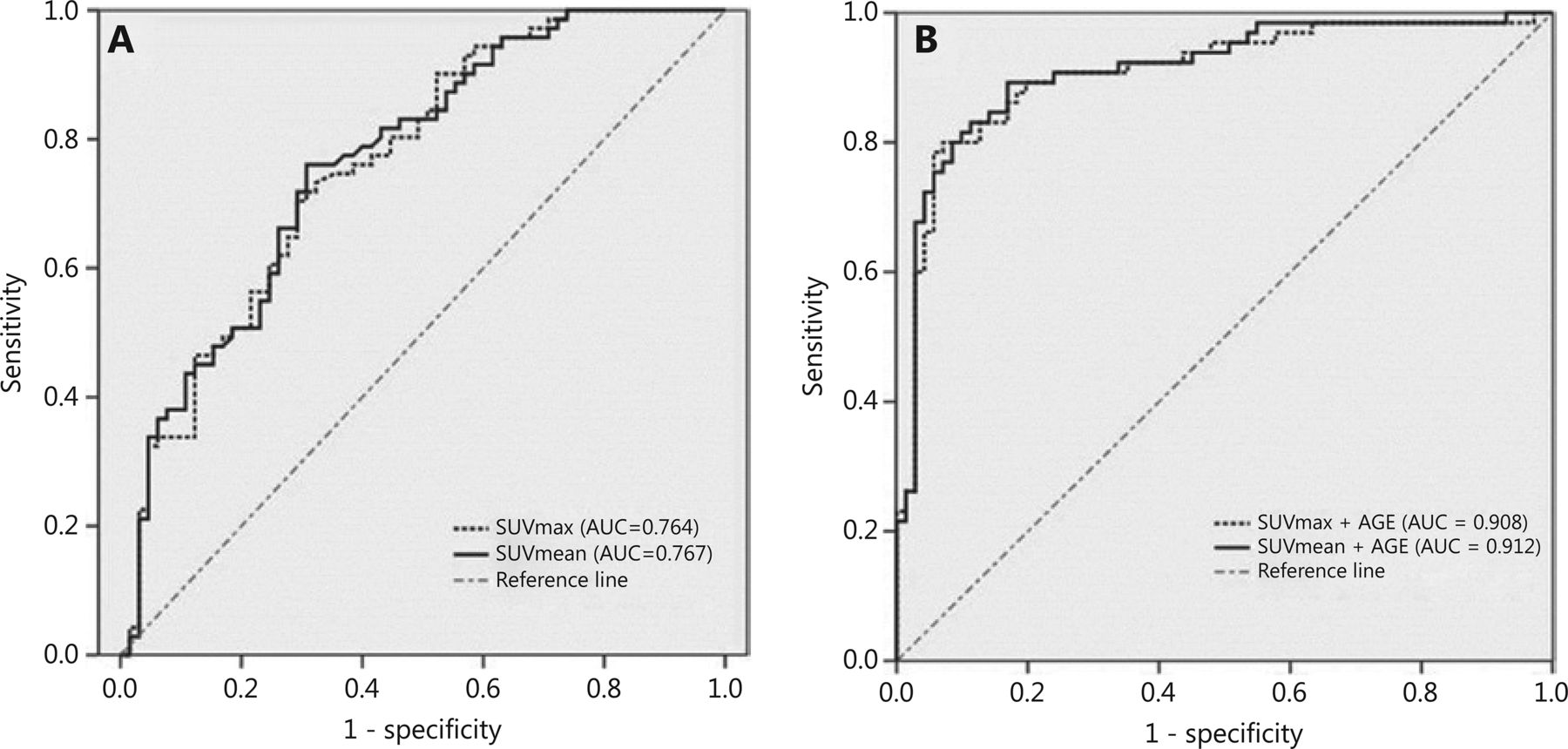
- Figure 5
ROC curve and area under the curve of the maximum standard uptake value and mean standard uptake value in differentiating patients with thymic epithelial tumors from patients with primary mediastinal lymphomas in the pre-vascular compartment of the mediastinum (A). The areas under the curve were significantly greater when age was involved as an index (B) in differentiating patients with thymic epithelial tumors from patients with primary mediastinal lymphomas.


Tables
- Table 1
Demographics of patients diagnosed with primary mediastinal lymphoma and thymic epithelial tumors in this study
Pathological subtypes N Age Age range Male (%) Primary mediastinal lymphoma 71 (52.2%) 30.25 ± 14.44 3–70 22 (31.0) Classic Hodgkin lymphoma 26 26.19 ± 9.82 6–50 23.1 Diffuse large B-cell lymphoma 30 36.43 ± 14.1 15–70 30.0 T-lymphoblastic lymphoma 9 19.33 ± 16.9 3–46 55.6 Other types 6 33.33 ± 16.86 13–59 33.3 Thymic epithelial tumors 65 (47.8%) 54.23 ± 15.16 12–77 40 (61.5) Thymoma_A 4 64.75 ± 7.41 55–73 50.0 Thymoma_AB 4 57.00 ± 9.13 46–68 50.0 Thymoma_B1 8 53.88 ± 12.51 41–76 62.5 Thymoma_B2 6 52.5 ± 21.67 16–72 66.7 Thymoma_B3 4 48.75 ± 13.3 33–64 75.0 Thymic carcinomas 39 53.77 ± 15.97 12–77 61.5 Squamous cell carcinoma 26 55.19 ± 15.48 12–76 61.5 Neuroendocrine carcinoma 8 58.25 ± 17.43 26–77 62.5 Adenocarcinoma/sarcoma 5 39.2 ± 8.58 24–45 60.0 Total 136 41.57 ± 19.12 3–77 62 (45.6) - Table 2
Group comparison between the primary mediastinal lymphoma and thymic epithelial tumors
Primary mediastinal lymphoma Thymic epithelial tumors Group comparison χ2 P Age 55.19 < 0.001** < 40 54 8 ≥ 40 17 57 Sex 12.68 < 0.001** Male 22 40 Female 49 25 Malignancy 33.21 < 0.001** Malignant 71 40 Benign 0 25 SUVmax 16.55 ± 6.38 10.64 ± 6.16 28.18 < 0.001** < 13.7 27 48 17.47 < 0.001** ≥ 13.7 44 17 SUVmean 9.80 ± 3.93 6.11 ± 3.55 28.93 < 0.001** < 8.0 29 49 16.43 < 0.001** ≥ 8.0 42 16 MTV (cm3) 143.98 ± 149.93 84.99 ± 130.10 5.74 < 0.05* < 115.8 39 53 10.9 = 0.001** ≥ 115.8 32 12 TLG (g/mL cm3) 1546.1 ± 1838.69 641.78 ± 1381.45 14.55 < 0.001** < 1113.9 42 57 13.85 < 0.001** ≥ 1113.9 29 8 SUVmax, maximum standardized uptake value; SUVmean, mean standardized uptake value; MTV, metabolic tumor volume; TLG, total lesion glycolysis; * and ** represent significant differences of P < 0.05 and P < 0.01, respectively.
- Table 3
Comparisons of metabolic and volumetric parameters in groups of patients with primary mediastinal lymphoma and thymic epithelial tumors
Pathological subtypes SUVmax SUVmean MTV TLG Total 13.72 ± 6.92 8.03 ± 4.17 115.79 ± 143.37 1113.95 ± 1692.13 Primary mediastinal lymphoma 16.55 ± 6.38 9.80 ± 3.93 143.98 ± 149.93 1546.15 ± 1838.69 Classic Hodgkin lymphoma 14.05 ± 5.27 8.51 ± 3.53 73.4 ± 101.75 669.34 ± 888.29 Diffuse large B-cell lymphoma 20.81 ± 5.69 12.28 ± 3.44 180.92 ± 143.32 2327.82 ± 2082.62 T-lymphoblastic lymphoma 11.94 ± 3.39 6.87 ± 1.95 188.06 ± 134.80 1459.41 ± 1387.87 Other types 12.99 ± 5.53 7.32 ± 3.25 199.05 ± 271.13 1567.05 ± 2733.1 χ2, P 23.34, < 0.001 22.47, < 0.001 13.15, 0.004 13.96, 0.003 Thymic epithelial tumors 10.64 ± 6.16 6.11 ± 3.55 84.99 ± 130.10 641.78 ± 1381.45 Thymoma_A 6.37 ± 0.86 3.76 ± 0.52 15.65 ± 7.77 56.34 ± 24.43 Thymoma_AB 5.37 ± 2.00 3.14 ± 1.28 52.94 ± 36.31 136.72 ± 53.78 Thymoma_B1 6.16 ± 2.61 3.59 ± 1.71 39.21 ± 22.59 122.32 ± 58.77 Thymoma_B2 8.65 ± 3.85 5.07 ± 2.30 43.29 ± 21.51 210.08 ± 137.81 Thymoma_B3 9.91 ± 8.48 5.97 ± 5.37 59.60 ± 38.04 317.35 ± 253.83 Thymic carcinomas 12.91 ± 6.29 7.35 ± 3.60 113.79 ± 160.87 959.87 ± 1715.11 Squamous cell carcinoma 14.22 ± 6.68 8.25 ± 3.86 99.86 ± 121.29 1005.18 ± 1872.20 Neuroendocrine carcinoma 9.99 ± 3.94 5.86 ± 2.34 184.24 ± 278.80 1168.21 ± 1754.16 Adenocarcinoma/sarcoma 10.78 ± 5.98 5.04 ± 2.06 73.54 ± 68.25 391.46 ± 386.17 χ2, P 22.51, 0.002 23.05, 0.002 11.58, > 0.05 20.61, 0.004 SUVmax, maximum standardized uptake value; SUVmean, mean standardized uptake value; MTV, metabolic tumor volume; TLG, total lesion glycolysis.
- Table 4
Diagnostic ability of metabolic and volumetric parameters of PET/CT in differentiating patients with primary mediastinal lymphoma versus thymic epithelial tumors
Cut-off values Sensitivity (%) Specificity (%) Accuracy (%) AUC (95% CI) SUVmax 12.3 70.4 70.8 70.6 0.764 (0.685–0.843) SUVmean 6.9 76.1 69.3 72.8 0.767 (0.688–0.847) TLG (g/mL cm3) 350.3 70.4 63.1 69.0 0.690 (0.599–0.780) MTV (cm3) 106 46.5 81.5 66.1 0.619 (0.524–0.715) SUVmax + TLG / 66.2 73.2 / 0.768 (0.689–0.847) SUVmax + age / 80.0 93.0 / 0.908 (0.855–0.961) SUVmean + age / 83.1 88.7 / 0.912 (0.861–0.964) SUVmax, maximum standardized uptake value; SUVmean, mean standardized uptake value; MTV, metabolic tumor volume; TLG, total lesion glycolysis.
In this issue


Jump to section
Related Articles
Cited By...
- No citing articles found.