

Abstract
OBJECTIVE To investigate the available parameters in gynecological screening for cervical lesions by liquid-based cytology technology (ThinPrep Cytology Test, TCT) and The Bethesda System (TBS), also with computer image analysis.
METHODS With application of the image analysis system, all grades of cervical lesion cells were detected quantitatively and sorted in atypical squamous cells of undetermined significance (ASCUS), atypical squamous cells-cannot exclude HSIL (ASC-H), low-grade squamous intraepithelial lesion (LSIL), high-grade squamous intraepithelial lesion (HSIL) and cervical squamous cell carcinoma (SCC) with the mean optical density (MOD), average grey (AG), positive units (PU), and nucleus to cytoplasmic ratio (N: C). Differences between each group of cells were compared and analyzed statistically.
RESULTS Apart from four stereologic parameters in LSIL and HSIL groups there were no differences among them, in the other groups, there was statistically significant in differences between MOD, AG and PU values. Differences between them in the ratio of nucleus to cytoplasm were highly statistically significant.
CONCLUSION Stereological indexes may serve as a screening tool for cervical lesions. The image analysis system is expected to become a new means of cytological assisted diagnosis.
keywords
Introduction
Cervical carcinoma is one of common gynecologic tumors. Early screening and intervention can prevent cervical cancer occurring[1]. Although manual checking of conventional cervical smears has been used for decades to screen for cervical cancer and precancerous cells. In many countries liquid based cytology is replacing conventional cytology. The potential advantages of liquid based cytology are simultaneous testing (including testing for human papilloma virus); faster reading times; and the cost savings due to automation[2]. The Bethesda System (TBS) and ThinPrep Cytology Test (TCT) are both liquid based cytologies which continue to be widely used, because they can increase the examination and accuracy of cervical lesions significantly[3,4]. The ThinPrep Imager system (Imager), a computerized system for reading slides, is a new technology applied to liquid based cytology. It has successfully combined modern computer technology and image analysis with the unique human interpretive skills of the cytotechnologist and pathologist. The ThinPrep Imaging System was approved by the US Food and Drug Administration in June of 2003 and has been widely adopted primarily[5-8]. The Imager has been utilized rarely in China due to the high cost. However, more recently the image analysis system has become increasingly common in clinical pathological practice. By means of the image analysis system and morphologic parameters, this study investigated available indexes for screening and diagnosis of cervical lesions. It was a preliminary attempt at automatic recognition by a computer in reporting TCT smears.
Materials and Methods
Materials
A Total of 296 cases of cervical lesion smears were selected from 2008 and 2009 archival files in the Department of Pathology of the First Affiliated Hospital of Zhengzhou University. These specimens were processed by the THINPREP 2000 Processor (Boxborough Cytyc Co., MA. USA). The specimens were made up of 2 cm diameter thin smears by the system programming, fixed by 95% ethanol, dyed with the Pap method, transpired in xylene, and sealed up with neutral balsam in that order. They include 93 cases of atypical squamous cells of undetermined signification (ASC-US), 58 cases of atypical squamous cells-cannot exclude HSIL (ASC-H), 55 cases of low-grade squamous intraepithelial lesion (LSIL), 51 cases of high-grade squamous intraepithelial lesion (HSIL), and 39 cases of cervical squamous cell carcinoma (SCC). High Resolution Pathological Image Analysis System (HPIAS-1000) was manufactured by the Wuhan Champion Image Engineering Company of Tongji Medical University. The HPIAS-1000 system mainly consists of JVC TK-C1381 (Japan) camera and PVPCI-M Capture VxD 1.0c (Microview, Beijing) capturing card inside of the computer.
Methods
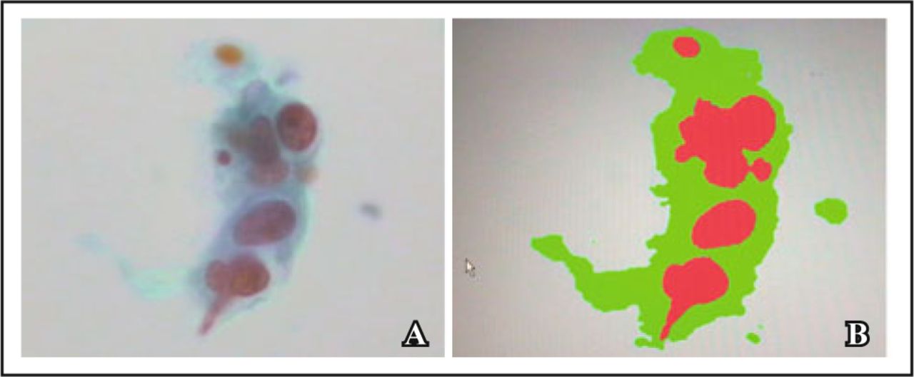
The HPIAS-1000 image analysis system was used to measure the values of these parameters with × 400 on the OLYMPUS CX-30 multifunctional microscope. The normal cervical epithelium nucleus, ASC-US, ASC-H, LSIL, HSIL, and SCC cells were surveyed for mean optical density (MOD), average grey (AG), positive unit (PU) of nucleus and nuclei-cytoplasm ratio (N:C). There were many stereologic parameters provided by the HPIAS-1000, such as MOD, PU, N:C, AG and so on. Theses indexes were achieved through the capturing card inside of the computer and special software. The greater value of AG is represented by the nucleus staining light. The greater value of PU and MOD is shown when the nucleus dyes dark[9-11]. Small cells, such as lymphocytes and epithelial cells should be avoided when measuring N:C ratio value. 5 typical lesion fields were selected for each case’s smear. More than 500 cells were searched for each kind of cervical lesion (Fig. 1).

A, Expression of HSIL cell with Papanicolaou smear (× 400); B, HSIL cells image of two-value segmentation with the image analysis system.
Statistical analysis
All testing values were presented as mean ± SD. Statistical significance in differences between the groups was assessed by Student’s t-test using SPSS 11.5.0 (SPSS Inc, 1989-2002) software. The results were considered statistically significant if the P value was < 0.05 or < 0.01. P < 0.01 was highly statistically significant.
Results
MOD, PU and N:C values were higher compared to normal epithelium cell, ASC-US, ASC-H, LSIL, HSIL and SCC. However, AG values were lower. Except for the 4 stereological parameters in LSIL and HSIL group, there were no differences among them (P > 0.05). In comparison between the other two groups, there was statistical significance (P < 0.05) in the differences between MOD, AG and PU values. Differences between them in the ratio of nucleus to cytoplasm were highly statistically significant (P < 0.01) (Table 1).
Stereological features at all levels of the cervix lesions ( ).
).
Discussion
Cancer of the cervix is a major threat to women’s health at present. The key to prevention and treatment of cervical carcinoma is cervical screening for early stage lesions being done effectively. Cervical cytology test has become an important means of general survey and a preferred method[12-15]. ThinPrep liquid-based cytology preparation has changed the way of doing smear from the conventional way. Samples are fixed in time and blood, mucus and unstructured pieces eliminated from the specimen. Thin layer smear is carried out in this way so the smear background and cell structure can be displayed clearly. In doing it this way the satisfaction of specimen can be increased. It was found to be good for recognition and diagnosis of cells. The rate of detection of lesions increased significantly[16-19].
Traditional cytological diagnosis depends on the diagnosing doctor’s professional experience. The diagnosis is qualitative analysis artificially and arbitrarily. The same cases may be diagnosed differently by different observers, even the same observer can diagnoses differently at different times. Traditional cytological diagnosis is lacking in objective and quantitative indicators. Applications of image analyzer and morphometric progressions have led to automatic identification of the abnormal cells by a computer system[20].
In the current study, automatic image analyzer and stereological generic parameters were utilized for quantitative detection of all kinds of abnormal cervical cells. MOD, PU and N:C values were higher than normal epithelium cell, ASC-US, ASC-H, LSIL, HSIL and SCC. AG values were lower. The 4 stereological parameters in LSIL and HSIL groups had no difference among them. In comparison between the other two groups, there was statistical significance in the differences between MOD, AG and PU values. Differences between them in N:C were highly statistically significant.
In The Bethesda System, LSIL and HSIL groups were used to describe the degree of atypia of cells in cervical lesion. It is difficult to distinguish them clearly. They were all designated to receive vaginoscopy biopsy and/or cervical conization after this. The rules that cytopathologists diagnose them with may be completely different. Although the ThinPrep Imaging System has been shown to considerably improve the sensitivity of cytology test, the key work is done by manual screening.
The current study shows that the Stereology index may serve as a measurement of cervical squamous lesions when used for screening in clinical tests and experimental studies[21]. Thus this image analysis system is expected to become a new auxiliary means of cytological diagnosis[22-23]. Overall, this technology will make a significant contribution to cervical cancer screening and patient management[24-25].
Conflict of interest statement
No potential conflicts of interest were disclosed.
Footnotes
This work was supported by a grant from the Natural Science Foundation of Henan Province, China (No.102300410078).
- Received January 15, 2010.
- Accepted October 10, 2010.
- Copyright © 2010 by Tianjin Medical University Cancer Institute & Hospital and Springer
References
In this issue


Jump to section
Related Articles
Cited By...
- No citing articles found.